Spalteholz HANDATLAS DER ANATOMIE DES MENSCHEN VON WERNER SPALTEHOLZ
メニューは解剖学(TA)にリンクしてあります。図の番号をクリックすると下記の説明へ、右側の用語をクリックすると解剖学(TA)にジャンプします。
346


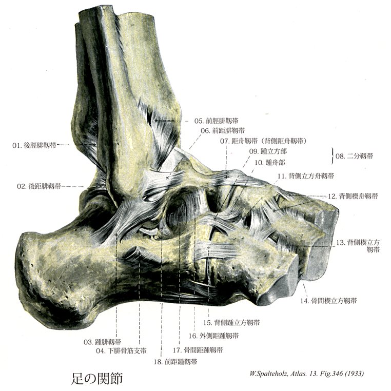
- 346_00【Joints of foot足の関節;足関節 Articulationes pedis】
→(距腿関節をも含めて、足根骨、中足骨および足の指骨の間に生ずるすべての関節を総称していう。狭義では距腿関節のみを指す。(解剖学辞典:河西達夫))
- 346_01【Posterior tibiofibular ligament後脛腓靱帯;後外果靱帯 Ligamentum tibiofibulare posterius; Ligamentum malleoli lateralis posterius】 Bands that pass on the posterior aspect of the inferior tibiofibular joint, connecting the end of the fibula with the lateral malleolus and stabilizing the malleolar mortise.
→(脛骨と腓骨の骨幹靱帯の後面には脛骨下端から腓骨下端に向かって外下方に走る後頚腓骨靱帯があって結合を強める。)
- 346_02【Posterior talofibular ligament後距腓靱帯;後腓距靱帯 Ligamentum talofibulare posterius; Ligamentum fibulotalare posterius】 Ligament that originates from the malleolar fossa of the lateral malleolus and inserts on the lateral tubercle of the talus.
→(後距腓靱帯は外果窩の底から起こり後内方に向かい、距骨後突起の外側結節に着く。)
- 346_03【Calcaneofibular ligament踵腓靱帯;腓踵靱帯 Ligamentum calcaneofibulare; Ligamentum fibulocalcaneare】 Ligament that passes from the tip of the lateral malleolus obliquely and posteriorly to the calcaneus.
→(踵腓靱帯は外果の下縁から起こり、距骨下関節の表面を越えて下方、やや後方に分散して距骨の外側面に着く。)
- 346_04【Inferior fibular retinaculum; Inferior peroneal retinaculum下腓骨筋支帯;遠位腓骨筋支帯 Retinaculum musculorum fibularium inferius; Retinaculum musculorum peroneorum inferius; Retinaculum musculorum fibularium distale】 Lower retinaculum that holds the peroneus muscles in place. It passes from the extensor retinaculum to the lateral surface of the calcaneus. One band passes to the fibular trochlea, dividing the peroneus brevis and peroneus longus muscles overlying it. It strengthens the dorsal fascia of the foot.
→(下腓骨筋支帯は下伸筋支帯の外側脚につづいて踵骨外側面から踵骨隆起外側面下部に至る。)
- 346_05【Anterior tibiofibular ligament前脛腓靱帯;前外果靱帯 Ligamentum tibiofibulare anterius; Ligamentum malleoli lateralis anterius】 Ligamentous bands that connect the anterior aspect of the end of the fibula with the lateral malleolus, stabilizing the malleolar mortise.
→(脛骨と腓骨の骨幹靱帯の前面には脛骨下端から腓骨下端に向かって外下方に走る前頚腓骨靱帯があって結合を強める。)
- 346_06【Anterior talofibular ligament前距腓靱帯;前腓距靱帯 Ligamentum talofibulare anterius】 Ligament that passes from the lateral malleolus to the lateral surface of the neck of the talus.
→(前距腓靱帯は外下の前縁から起こり、前内方に向かって距骨頚の外側部に着く。)
- 346_07【Talonavicular ligament距舟靱帯;背側距舟靱帯 Ligamentum talonaviculare; Ligamentum talonaviculare dorsale】 Dorsal band that passes from the head of the talus to the navicular.
→(距舟靱帯は距踵舟関節距舟部の関節包の一部で、距骨頚と舟状骨の間の背面に張る。)
- 346_08Chopart's ligament【Bifurcate ligament二分靱帯 Ligamentum bifurcatum】 Y-shaped ligament in front of the tarsal sinus on the dorsum of the foot. It passes anteriorly from the calcaneus and consists of the following two parts.
→(二分靱帯は踵骨背面の前内側部(足根洞の底の前方部)から起こって前方に向かう強い靱帯で、内外2部に分かれる。内側部は距舟靱帯で、距踵舟関節包の内側部となる。外側部は踵立方靱帯といい、踵立方関節の背面内側部を強める。)
- 346_09【Calcaneocuboid ligament踵立方靱帯;踵立方部(二分靱帯の) Ligamentum calcaneocuboideum; Pars calcaneocuboidea】 Portion of the bifurcate ligament that extends from the calcaneus and nearly reaches the middle of the cuboid.
→(二分靱帯の外側部は踵立方靱帯といい、踵立方関節の背面内側部を強める。)
- 346_10【Calcaneonavicular ligament踵舟靱帯;踵舟部(二分靱帯の) Ligamentum calcaneonaviculare; Pars calcaneonavicularis】 Medial portion of the bifurcate ligament that passes from the calcaneus to the navicular.
→(踵舟靱帯は二分靱帯の内側部で、踵骨背側面の前内側部を起点として前走し舟状骨に付き、距踵舟関節包の内側の部分を作る。)
- 346_11【Dorsal cuboideonavicular ligament背側立方舟靱帯 Ligamentum cuboideonaviculare dorsale】 Band that connects the cuboid and navicular.
→(背側立方舟靱帯は立方骨と舟状骨の背面を結ぶ。)
- 346_12【Dorsal cuneonavicular ligament背側楔舟靱帯;背側舟楔靱帯 Ligamenta cuneonavicularia dorsalia; Ligamenta navicularicuneiformia dorsalia】 Broad bands on the dorsum of the foot that connect the navicular with the three cuneiform bones.
→(背側楔舟靱帯は舟状骨と内側・中間・外側楔状骨の背面を結び、楔舟関節包の背部を作る。)
- 346_13【Dorsal cuneocuboid ligament背側楔立方靱帯 Ligamentum cuneocuboideum dorsale】 Dorsal band that extends from the lateral cuneiform to the cuboid.
→(背側楔立方靱帯は外側楔状骨と立方骨の背面を結び、前者の外側縁を中心として放散する走行を示す。)
- 346_14【Cuneocuboid interosseous ligament骨間楔立方靱帯 Ligamentum cuneocuboideum interosseum】 Tough band that passes from the lateral cuneiform to the cuboid.
→(骨間楔立方靱帯は外側楔状骨と立方骨の対向する面を結ぶ。)
- 346_15【Dorsal calcaneocuboid ligament背側踵立方靱帯 Ligamentum calcaneocuboideum dorsale】 Moderately thickened portion of the joint capsule lateral to the bifurcate ligament.
→(背側踵立方靱帯は二分靱帯の外側関節包(踵立方靱帯)を補強する。)
- 346_16【Lateral talocalcaneal ligament外側距踵靱帯;腓側距踵靱帯 Ligamentum talocalcaneum laterale; Ligamentum talocalcaneum fibulare】 Ligament that passes from the trochlea of the talus to the lateral surface of the calcaneus. It is partly covered by the calcaneofibular ligament.
→(外側距踵靱帯は距骨の外側突起前部から出て下後方に向かい、踵骨の外側面に着く。)
- 346_17【Talocalcaneal interosseus ligament骨間距踵靱帯 Ligamentum talocalcaneum interosseum】 Strong bands of fibers in the tarsal sinus that divide the upper and lower parts of the ankle joint.
→(骨間距踵靱帯は距骨溝と踵骨溝が合して作る足根洞の内部で距骨と踵骨とを結ぶ板状の靱帯である。足根洞の内側半部は狭い管状で、ここにある靱帯は距踵舟関節包と距骨下関節包の一部である。外側半部は漏斗状に拡がり、ここに交叉性に線維が走る強い靱帯がある。足根洞の靱帯以外の部は脂肪組織でみたされている。)
- 346_18【Anterior talocalcaneal ligament前距踵靱帯 Ligamentum talocalcaneum anterius】
→()