Spalteholz HANDATLAS DER ANATOMIE DES MENSCHEN VON WERNER SPALTEHOLZ
メニューは解剖学(TA)にリンクしてあります。図の番号をクリックすると下記の説明へ、右側の用語をクリックすると解剖学(TA)にジャンプします。
348


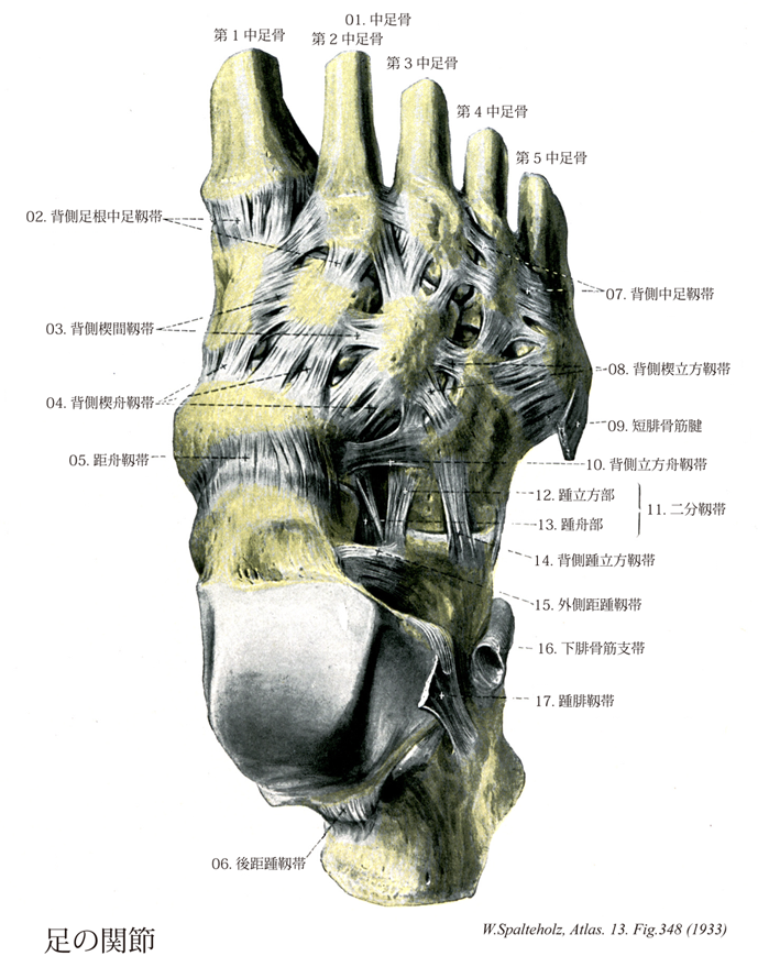
- 348_00【Joints of foot足の関節;足関節 Articulationes pedis】
→(距腿関節をも含めて、足根骨、中足骨および足の指骨の間に生ずるすべての関節を総称していう。狭義では距腿関節のみを指す。(解剖学辞典:河西達夫))
- 348_01【Metatarsals; Metatarsal bones [I-V]中足骨[1-5] Ossa metatarsi; Ossa metatarsalia [I-V]】 The five metatarsal bones.
→(中足の骨格で5個の長骨からなる。内側から順に第1・第2・第3・第4および第5中足骨という。第1・第2および第3中足骨はそれぞれ内側・中間および外側楔状骨の遠位に、第4および第5中足骨は立方骨の遠位にある。長さは中手骨より長い。第1中足骨が最も短く、第2中足骨が最も長い。第3・第4および第5中足骨の順に短くなる。おのおのの中足骨を近位端の底、中央部の体、遠位端の頭に分ける。底は太く厚く、第1・第2および第3中足骨にはそれぞれ内側・中間および外側楔状骨に対する、第4と第5中足骨には立方骨に対する関節面がある。第1中足骨を除く中足骨と足根骨の関節を連ねる線は、内側前方から外側後方へ走る。第2・第3・第4および第5中足骨底では相対する側面に関節面があるが、第1中足骨底にはない。また第2中足骨底の側面には内側および外側楔状骨に対する関節面がある。第1中足骨底の足底面に第1中足骨粗面がある。第5中足骨底の外側は強く張り出して第5中足骨粗面をなし、皮下で触知できる。体は不正三角柱で、第1中足骨では太いが、他は左右から圧迫された形をし、頭へいくほど細くなる。また長軸方向で背側に凸弯している。頭は側面を切り取った球状で、細い頚部がある。頭の足底面には内および外側に、それぞれちいさな隆起がある。)
- 348_01a【First metatarsal bone; 1st metatarsal bone第1中足骨 Os metatarsale I】
→()
- 348_01b【Second metatarsal bone; 2nd metatarsal bone第2中足骨 Os metatarsale II】
→()
- 348_01c【Third metatarsal bone; 3rd metatarsal bone第3中足骨 Os metatarsale III】
→()
- 348_01d【Fourth metatarsal bone; 4th metatarsal bone第4中足骨 Os metatarsale IV】
→()
- 348_01e【Fifth metatarsal bone; 5th metatarsal bone第5中足骨 Os metatarsale V】
→()
- 348_02【Dorsal tarsometatarsal ligaments背側足根中足靱帯 Ligamenta tarsometatarsalia dorsalia】 Ligaments on the dorsal side of the foot that connect the tarsus with the metatarsals.
→(背側足根中足靱帯は相対応する中足骨と遠位足根骨とを背面で結ぶ。)
- 348_03【Dorsal intercuneiform ligament背側楔間靱帯 Ligamenta intercuneiformia dorsalia】 Dorsal bands that connect the cuneiform bones.
→(背側楔間靱帯は各楔状骨間の背面を結ぶ。)
- 348_04【Dorsal cuneonavicular ligament背側楔舟靱帯;背側舟楔靱帯 Ligamenta cuneonavicularia dorsalia; Ligamenta navicularicuneiformia dorsalia】 Broad bands on the dorsum of the foot that connect the navicular with the three cuneiform bones.
→(背側楔舟靱帯は舟状骨と内側・中間・外側楔状骨の背面を結び、楔舟関節包の背部を作る。)
- 348_05【Talonavicular ligament距舟靱帯;背側距舟靱帯 Ligamentum talonaviculare; Ligamentum talonaviculare dorsale】 Dorsal band that passes from the head of the talus to the navicular.
→(距舟靱帯は距踵舟関節距舟部の関節包の一部で、距骨頚と舟状骨の間の背面に張る。)
- 348_06【Posterior talocalcaneal ligament後距踵靱帯 Ligamentum talocalcaneum posterius】 Band that passes from the posterior process of the talus to the calcaneus. It spans the groove for the tendon of the flexor hallucis longus.
→(距骨下関節の関節包の前壁は足根洞にある骨間距舟靱帯の一部と成る。後壁のうち、距骨後突起の内、外側結節から分かれて起こる線維束は後距踵靱帯といい、内側結節から起こる部は長母趾屈筋腱の表面を越える。)
- 348_07【Dorsal metatarsal ligaments背側中足靱帯;背側中足骨底靱帯 Ligamenta metatarsalia dorsalia; Ligamenta basium ossium metatarsi dorsalia】 Fibrous bands connecting the dorsal surfaces of the bases of the metatarsals.
→(背側中足靱帯は各中足骨底の背面を横に結ぶ。)
- 348_08【Dorsal cuneocuboid ligament背側楔立方靱帯 Ligamentum cuneocuboideum dorsale】 Dorsal band that extends from the lateral cuneiform to the cuboid.
→(背側楔立方靱帯は外側楔状骨と立方骨の背面を結び、前者の外側縁を中心として放散する走行を示す。)
- 348_09【Fibularis (peroneus) brevis tendon; Tendon of peroneus brevis muscle短腓骨筋腱 Tendo musculus peroneus brevis】
→()
- 348_10【Dorsal cuboideonavicular ligament背側立方舟靱帯 Ligamentum cuboideonaviculare dorsale】 Band that connects the cuboid and navicular.
→(背側立方舟靱帯は立方骨と舟状骨の背面を結ぶ。)
- 348_11Chopart's ligament【Bifurcate ligament二分靱帯 Ligamentum bifurcatum】 Y-shaped ligament in front of the tarsal sinus on the dorsum of the foot. It passes anteriorly from the calcaneus and consists of the following two parts.
→(二分靱帯は踵骨背面の前内側部(足根洞の底の前方部)から起こって前方に向かう強い靱帯で、内外2部に分かれる。内側部は距舟靱帯で、距踵舟関節包の内側部となる。外側部は踵立方靱帯といい、踵立方関節の背面内側部を強める。)
- 348_12【Calcaneocuboid ligament踵立方靱帯;踵立方部(二分靱帯の) Ligamentum calcaneocuboideum; Pars calcaneocuboidea】 Portion of the bifurcate ligament that extends from the calcaneus and nearly reaches the middle of the cuboid.
→(二分靱帯の外側部は踵立方靱帯といい、踵立方関節の背面内側部を強める。)
- 348_13【Calcaneonavicular ligament踵舟靱帯;踵舟部(二分靱帯の) Ligamentum calcaneonaviculare; Pars calcaneonavicularis】 Medial portion of the bifurcate ligament that passes from the calcaneus to the navicular.
→(踵舟靱帯は二分靱帯の内側部で、踵骨背側面の前内側部を起点として前走し舟状骨に付き、距踵舟関節包の内側の部分を作る。)
- 348_14【Dorsal calcaneocuboid ligament背側踵立方靱帯 Ligamentum calcaneocuboideum dorsale】 Moderately thickened portion of the joint capsule lateral to the bifurcate ligament.
→(背側踵立方靱帯は二分靱帯の外側関節包(踵立方靱帯)を補強する。)
- 348_15【Lateral talocalcaneal ligament外側距踵靱帯;腓側距踵靱帯 Ligamentum talocalcaneum laterale; Ligamentum talocalcaneum fibulare】 Ligament that passes from the trochlea of the talus to the lateral surface of the calcaneus. It is partly covered by the calcaneofibular ligament.
→(外側距踵靱帯は距骨の外側突起前部から出て下後方に向かい、踵骨の外側面に着く。)
- 348_16【Inferior fibular retinaculum; Inferior peroneal retinaculum下腓骨筋支帯;遠位腓骨筋支帯 Retinaculum musculorum fibularium inferius; Retinaculum musculorum peroneorum inferius; Retinaculum musculorum fibularium distale】 Lower retinaculum that holds the peroneus muscles in place. It passes from the extensor retinaculum to the lateral surface of the calcaneus. One band passes to the fibular trochlea, dividing the peroneus brevis and peroneus longus muscles overlying it. It strengthens the dorsal fascia of the foot.
→(下腓骨筋支帯は下伸筋支帯の外側脚につづいて踵骨外側面から踵骨隆起外側面下部に至る。)
- 348_17【Calcaneofibular ligament踵腓靱帯;腓踵靱帯 Ligamentum calcaneofibulare; Ligamentum fibulocalcaneare】 Ligament that passes from the tip of the lateral malleolus obliquely and posteriorly to the calcaneus.
→(踵腓靱帯は外果の下縁から起こり、距骨下関節の表面を越えて下方、やや後方に分散して距骨の外側面に着く。)