Spalteholz HANDATLAS DER ANATOMIE DES MENSCHEN VON WERNER SPALTEHOLZ
メニューは解剖学(TA)にリンクしてあります。図の番号をクリックすると下記の説明へ、右側の用語をクリックすると解剖学(TA)にジャンプします。
544 


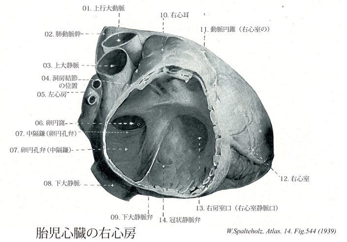
- 544_01【Ascending aorta上行大動脈;大動脈上行部 Pars ascendens aortae; Aorta ascendens】 Ascending part of the aorta up to its exit from the pericardium.
→(左心室からおこり、肺動脈幹の後ろを上行して大動脈弓にいたる5cmほどの部。基部の内腔は膨らんで大動脈洞(バルサルバ洞)をなし、ここから左右の冠状動脈が出る。(イラスト解剖学))
- 544_02【Pulmonary trunk; Pulmonary artery肺動脈幹;肺動脈 Truncus pulmonalis; Arteria pulmonarlis】 Arterial trunk that ascends in the pericardium. It divides into the right and left pulmonary arteries at the level of the reflection of the serous pericardium.
→(肺動脈幹は右心房と左右肺動脈の起始までの間で分岐前の肺動脈を明確にするために導入された。肺動脈幹は左の第3肋骨の胸骨付近の高さで右心部動脈円錐から起こる。長さは約5cmで心膜腔を通り、やや左側に向かい、頭背側で肺動脈幹のT字形の分岐となり、2本の肺動脈に分岐する。肺動脈幹の両側を2本の冠状動脈が走る。肺動脈幹の起始部は大動脈口の前左側に、中間部は上行大動脈の左側に、分岐部は大動脈弓の凹部に位置する。)
- 544_03【Superior vena cava上大静脈 Vena cava superior; Vena cava cranialis】
→(上大静脈は上半身の血液を集める静脈で、上縦隔の中で左右の腕頭静脈が合してはじまり、途中で奇静脈を受け入れながら上行大動脈の右側を下行して右心房にそそぐ。)
- 544_04Keith-Flack, Node of; Koch's node【Sinu-atrial node; SA node; node of Keith and Flack洞房結節;洞結節;キース・フラックの結節 Nodus sinuatrialis】 Bandlike specialized muscle tissue in front of the entrance of the superior vena cava that functions as the primary center for generating cardiac impulses.
→(キース・フラックの結節ともよばれる。洞結節は上大静脈の開口部の前方に帯状にある特殊伸筋組織で、心拍動をきめる最初の刺激発生中心として働く。刺激激伝導系のペースメーカーをなす洞房結節のこと。洞房結節に規則正しく発生する興奮のほかに、何らかの理由で刺激伝導系の一部に興奮が発生すると、余分に収縮が起こる。このような余分に起こる収縮のことを期外収縮という。イギリスの解剖学・人類学者Sir Arthur Keith (1866-1953)と、同じくイギリスの生理学者Martin Flack (1882-1931)により、1970年にモグラの心臓で発見された。)
- 544_05【Left atrium左心房 Atrium cordis sinistrum; Atrium sinistrum】
→(左心房は心臓の後上部にあって、後面をつくっている。左心房は右心房よりもやや小さいが、壁はやや厚い。左心房の後壁の上部に、左右両肺からそれぞれ2本ずつ、前部で4本の肺静脈が開口している。左心房は前下方で房室口によって左心室に通じる。)
- 544_06【Fossa ovalis; Oval fossa卵円窩 Fossa ovalis】 Depression in the interatrial septum, which represents the closed foramen ovale (open during fetal development).
→(卵円窩は心房中隔で、胎生時の卵円孔の跡の凹み。)
- 544_07【Valve of foramen ovale卵円孔弁;中隔鎌 Valvula foraminis ovalis; Falx septi (atriorum)】 Openings of pulmonary veins into the left atrium.
→(卵円孔弁は成人では閉じて中隔鎌とよばれる。一次中隔に由来する卵円窩の底。胎児では左心房の血流で押しのけられる。)
- 544_08【Inferior vena cava下大静脈 Vena cava inferior; Vena cava caudalis】 It arises at the union of the right and left common iliac veins, lies on the right side of the aorta, and opens into the right atrium of the heart.
→(下大静脈は下肢および骨盤と腹部の器官の大部分から血液を受ける本幹で、第5腰椎体の右側で左右の総腸骨静脈の合流として始まり、このあと脊柱に沿って大動脈の右側を上行、肝臓の後面をこれに接して通過し、第八胸椎の高さで横隔膜の大静脈孔を貫いて胸腔に入り、ただちに右心房にそそぐ。下大静脈に流入する枝には総腸骨静脈、下横隔静脈、第3・第4腰静脈、肝静脈、腎静脈、右副腎静脈、右精巣静脈、右卵巣静脈、蔓状静脈叢などがある)
- 544_09Eustachian valve【Valve of inferior vena cava下大静脈弁 Valvula venae cavae inferioris】 Semilunar fold in front of the opening of inferior vena cava. During fetal development it directs blood toward the foramen ovale.
→(オイスタヒ弁とも呼ばれる。右心房内の下大静脈開口部前縁にみられる弁で、冠状静脈弁(テベシウス弁valve of Thebesius)とともに胎生期の静脈弁に由来するという。)
- 544_10【Right auricle of atrium右心耳 Auricula atrii dextra; Auricula dextra cordis】 Outpouching of the right atrium.
→(右心耳は右心房の中空指状突出。心耳の壁はうすく内面に櫛状筋が発達している。)
- 544_11【Conus arteriosus; Infundibulum of right ventricle動脈円錐;漏斗(右心室の) Conus arteriosus; Infundibulum】 Funnelshaped smooth-walled outflow tract leading to the pulmonary trunk.
→(右心室の動脈円錐は肺動脈管へ開く前の、漏斗状で平滑な壁からなる流出路。発生学的には心球にあたる。)
- 544_12【Right ventricle右心室 Ventriculus dexter】
→(右心室は心臓の最下位部を占め、後上方にある右房室口で右心房と交通し、前上方にある肺動脈口で肺静脈に連なる。)
- 544_13【Right atrioventricular orifice; Right atrioventricular opening右房室口;右心室静脈口 Ostium atrioventriculare dextrum; Ostium venosum ventriculi dextri】 Openings between the atria and ventricles.
→(右房室口は右心房と右心室の間に開く。右房室口は輪状の線維性結合織(線維輪)で囲まれ、弁をもつ。)
- 544_14Thebesian valve【Valve of coronary sinus冠状静脈弁;冠状静脈洞弁 Valvula sinus coronarii】 Semilunar fold in front of the opening of coronary sinus.
→(テベシウス弁ともよばれる。冠状静脈弁は冠状静脈洞が右心房に開口する部にみられる心内膜のヒダ。オイスタヒ弁(下大静脈弁)とともに胎生期の静脈弁に由来する。)