Spalteholz HANDATLAS DER ANATOMIE DES MENSCHEN VON WERNER SPALTEHOLZ
メニューは解剖学(TA)にリンクしてあります。図の番号をクリックすると下記の説明へ、右側の用語をクリックすると解剖学(TA)にジャンプします。
584 


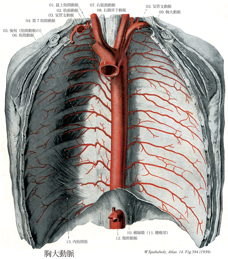
- 584_01【Supreme intercostal artery; Highest intercostal artery最上肋間動脈 Arteria intercostalis suprema】 Common trunk for the first two intercostal arteries.
→(最上肋間動脈は肋骨頚の前を下行枝、第1および第2肋間動脈となる。他の肋間動脈と同様に、背枝と脊髄枝を分枝する。)
- 584_02【Costocervical trunk肋頚動脈 Truncus costocervicalis】 Origin: posterior wall of subclavian artery, behind the anterior scalene muscle. Trunk of deep cervical artery and supreme intercostal artery.
→(肋頚動脈は鎖骨下動脈の後側でおこり、まもなく2枝に分かれる。①深頚動脈、②最上肋間動脈)
- 584_03【Bronchial branches of thoracic aorta; Bronchial arteries気管支動脈;気管支枝(胸大動脈の) Rami bronchiales (Aorta thoracica)】 Their origin is highly variable, often at the level of the tracheal bifurcation. They ramify along the bronchi, extend to the bronchioles and supply their walls and the connective-tissue septa of the lungs. They form anastomoses with branches of the pulmonary artery.
→(気管支動脈は2~3本で、大胸動脈や付近の動脈から起こり、肺門から肺に入る。気管支およびその分枝に沿って走り、呼吸細気管支にいたるまでの気道、小葉間結合組織、臓側胸膜に分布する。)
- 584_04【Seventh posterior intercostal artery; 7th intercostal artery第7肋間動脈 Arteria intercostalis posterior VII】
→()
- 584_05【Dorsal branch of posterior intercostal artery後枝;背側枝(肋間動脈の) Ramus dorsalis (Arteria intercostalis posterioris)】 Branch that passes posteriorly between the vertebral bodies and superior costotransverse ligament. It supplies the muscles and skin of back as well as the spinal cord and its meninges.
→(肋間動脈の背枝は1分節以下の肋骨頚の上を通って背側に向かい、次いで胸神経後枝と共に1分節以下の椎骨の横突起の上をこえて背部に出て、内側皮枝と外側皮枝に分かれ背部の皮膚に分布する。また途中、固有背筋に枝を与える。脊髄枝は、椎間孔のところで分岐して脊柱管に入り、脊髄とその皮膜へ分布する。)
- 584_06【Posterior intercostal arteries; Intercostal arteries肋間動脈;後肋間動脈(第3~第11) Arteriae intercostales posteriores】 Paired tributaries arising from the posterior wall of the aorta, supplying the third through eleventh intercostal spaces.
→(現在の学名ではintercostalesの後にposterioresを付して命名されているが、日本名ではたんに肋間動脈とよぶ。古い学名BNAやINAではArteriae intercostalesであった。Posterioresを付した理由は、内胸動脈より分枝する前肋間枝(Rami intercostales anteriores)と対比した物である。したがって胸大動脈の枝としての肋間動脈は、第三から第十一にいたる9対の肋間動脈を指す。左右とも胸大動脈の背外側面より分岐するが、大動脈が脊柱の左側にあるため、右肋間動脈は左よりも長く、また分岐後ただちに脊柱の前面を横切ることになる。左右とも交感神経幹の背側を通ってそれぞれの肋間腔に入ると、ただちに背枝を分岐し、本幹はそのまま側方へ走って、はじめ側壁胸膜下でこれと内肋間膜の間を走るが、肋骨の外側部のあたりでは、最内肋間筋と内肋間筋の間を走る。各肋間隙を通るときは、同じ番号の肋間の下縁(肋骨溝)に沿って肋間静脈と肋間神経にはさまれて走るが、このときは前者は動脈の頭側に、後者は尾側にあるのが原則である。肋骨の外1/3と前1/3の境界のあたりで、内胸動脈の枝である前肋間枝と吻合しておわるが、下位の肋間動脈の場合は、その末梢は腹壁に入り、腹横筋と内腹斜筋の間を前進して上腹壁動脈や肋下動脈と吻合する。)
- 584_07【Right common carotid artery右総頚動脈 Arteria carotis communis dextra】
→(総頚動脈は頭部に血液を送る血管の主幹。右は腕頭動脈の枝、左は大動脈弓の上行部より出る。そのため左総頚動脈は右のものよりも4~5cm長い。総頚動脈は枝を出さず、気管・喉頭の両側を上行し、甲状軟骨上縁の高さで音叉のような形をなし内・外頚動脈に分かれる。分岐部の後側には頚動脈小体が存在する。また分岐部のないし内頚動脈始部の壁はやや薄く膨隆しており(頚動脈洞)、舌咽神経の枝を介し血圧を感受するという。)
- 584_07a【Common carotid artery総頚動脈 Arteria carotis communis】 Artery of the neck without any branches. It runs on both sides of the trachea and larynx and passes deep to the sternocleidomastoid. It arises on the right from the brachiocephalic trunk and on the left from the aortic arch.
→(総頚動脈は頭部に血液を送る血管の主幹。右は腕頭動脈の枝、左は大動脈弓の上行部より出る。そのため左総頚動脈は右のものよりも4~5cm長い。総頚動脈は枝を出さず、気管・喉頭の両側を上行し、甲状軟骨上縁の高さで音叉のような形をなし内・外頚動脈に分かれる。分岐部の後側には頚動脈小体が存在する。また分岐部のないし内頚動脈始部の壁はやや薄く膨隆しており(頚動脈洞)、舌咽神経の枝を介し血圧を感受するという。)
- 584_08【Right subclavian artery右鎖骨下動脈 Arteria subclavia dextra】
→()
- 584_08a【Subclavian artery鎖骨下動脈 Arteria subclavia】 Artery that passes with the roots of brachial plexus between the anterior and middle scalene muscles through the scalene space, over the first rib in the groove for the subclavian artery. From the lateral border of the first rib, it continues as the axillary artery.
→(鎖骨下動脈は上肢の主幹動脈の根部をなし、右側は腕頭動脈から、左側は大動脈弓からそれぞれ分かれてはじまり、前斜角筋の後方を通って第1肋骨外側縁で腋窩動脈につづく。胸・頚・上肢移行部の動脈として、多彩な分枝と変異に富むことを特徴とする。分枝はつぎの通りである。椎骨動脈、内胸動脈、甲状頚動脈、肋頚動脈、下行肩甲動脈に分枝し、第一肋骨を越えたところで腋窩動脈となる。)
- 584_09【Thoracic aorta胸大動脈;大動脈胸部 Pars thoracica aortae; Aorta thoracica】 Part of the aorta descending to the aortic hiatus of the diaphragm at the level of the twelfth thoracic vertebra.
→(胸大動脈は、大動脈弓の延長である。第4胸椎体の下縁の左側で始まり、第5から第12胸椎の左側で後縦隔を下行する。下行しながら、正中面に近付き、食道と脊柱の左側に沿って走るが、食道の後方、脊柱の前を走るようになる。胸大動脈は横隔膜を貫いた直後に腹大動脈という名前に変わる。胸大動脈と腹大動脈とを総称して下行大動脈という。)
- 584_10【Diaphragm横隔膜 Diaphragma】 Dome-shaped musculofibrous septum dividing thoracic and abdominal cavities. I: Phrenic nerve.
→(横隔膜は胸腔と腹腔との境を作る膜状筋で、胸郭下口の周りから起こる。この起始部を腰椎部、肋骨部、胸骨部の3部に分ける。これらの部から出る筋束は全体として円蓋のように胸腔に盛り上がって集まり、中央部の腱膜につく。これを腱中心という。横隔膜の上面は胸内筋膜および胸膜と心膜、下面は横隔膜筋膜(横筋筋膜の一部)および腹膜(肝臓その他の臓器が接する部分を除いて)被われる。ドーム状の横隔膜は胸腔の床および腹腔の天井となる。閉鎖した筋腱様のしきりは哺乳類の特質である。横隔膜は最重要な呼吸筋である。筋素材は系統発生的に第3~5の頚部筋節から由来し、頚神経叢からの横隔神経(C4(3,5))に支配される。筋性横隔膜は腰椎部、肋骨部、胸骨部から形成される。)
- 584_11【Lumbar part of diaphragm腰椎部(横隔膜の) Pars lumbalis (Diaphragmatis)】 Part of the diaphragm that arises from the lumbar vertebrae, intervertebral discs, and tendinous arches.
→(横隔膜の腰椎体からの起始は腰椎部と呼ばれ、左右の2脚からなる。左脚は第1~第3腰椎体から、右脚は第1~第4腰椎体から、椎体前面の前縦靱帯と密着して腱様に起こって上行している。)
- 584_12Haller's tripus【Coeliac trunk; Celiac trunk; Celiac artery腹腔動脈 Truncus coeliacus】 Frequently a common trunk of the left gastric, common hepatic, and splenic arteries at the level of the twelfth thoracic vertebra. The left gastric artery ca. also branch off of the aorta earlier.
→(腹腔動脈は横隔膜直下の腹大動脈より起こり、左胃動脈、総肝動脈、脾動脈の共通幹で、第十二胸椎の高さにある。)
- 584_13【Internal intercostal muscle内肋間筋 Musculi intercostales interni】 Muscles that extend between the intercostal spaces from the sternum to the costal angle, passing from anterosuperior to posteroinferior. Expiratory muscles; fixation of the ribs. I: Intercostal nerves.
→(内肋間筋は全肋間内で外肋間筋によって覆われている。この筋線維は(外肋間筋に対して約90°の方向を示すが)後下方から前上方に走行する(下方では明確な境界を示すことなく付着する内腹斜筋の筋線維と同様に)。内肋間筋は腹側では胸骨まで、背側では肋骨角にまで広がっているにすぎない。肋骨の背側端では、筋線維束は腱様の内肋間膜によって置換されている。内肋間筋の深層は肋間動静脈と神経によって分けられ、最内肋間筋となる。肋軟骨間に位置する内肋間筋の位置する内肋間筋の部分は軟骨間筋とも言われる。)
