Spalteholz HANDATLAS DER ANATOMIE DES MENSCHEN VON WERNER SPALTEHOLZ
メニューは解剖学(TA)にリンクしてあります。図の番号をクリックすると下記の説明へ、右側の用語をクリックすると解剖学(TA)にジャンプします。
640


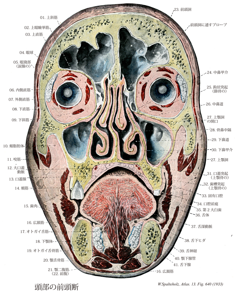
- 640_01【Superior oblique muscle上斜筋;上眼球斜筋 Musculus obliquus superior; Musculus obliquus bulbi superior】 o:Medial to the common tendinous ring on the body of sphenoid, i: After a hook-shaped course, obliquely behind the equator. Its tendon passes through the trochlea. Action: Abduction, intorsion, and depression of the eye. I: Trochlear nerve.
→(上斜筋は眼窩傍結合組織すなわち視神経鞘と(おもに)蝶形骨体の結合組織である総腱輪の内側から起こる。上斜筋は眼窩錐体の内側直近の上を前方に走行する。眼球の縁で上斜筋の丸みのある腱は結合組織性の吊り索(滑車)を通過し鋭角で後方に曲がる。さらに上斜筋の腱は上直筋の下でこれと交差し眼球上後側頭部の強膜に停止する。目の動き:視線を内側かつ下方に向ける。)
- 640_02【Levator palpebrae superioris muscle上眼瞼挙筋 Musculus levator palpebrae superioris】 o: Upper portion of optic canal and dural sheath of optic nerve. Its insertion tendon widens anteriorly and divides into a superior and an inferior layer. I: Oculomotor nerve.
→(上眼瞼挙筋は視神経管の縁の総腱輪の外側で視神経鞘から起こり、眼窩上壁のすぐ下で前頭神経の下を通り上眼瞼にいく。上眼瞼挙筋の腱は分離して上眼瞼挙筋浅板と上眼瞼挙筋深板に分かれる。前者は上眼瞼中を縁に向かって進み、後者は上瞼板筋の平滑筋細胞を伴って上眼瞼の瞼板に付く。下瞼板筋は下眼瞼板と下結膜円蓋の間の下眼瞼に存在する平滑筋層である。)
- 640_03【Superior rectus muscle上直筋;上眼球直筋 Musculus rectus superior; Musculus retus bulbi superior】 o: Common tendinous ring, i: Along an oblique line passing anterior to the equator, 7-8 mm behind the corneal margin. Action: Elevation and intorsion of superior pole. I: Oculomotor nerve.
→(上直筋は、眼球の上部を斜め外側に進んで眼球の周囲に達し、そこで角膜縁の後方約7-8mmの胸膜に停止腱が放射上に胸膜組織と絡まるように停止する。目の動き:視線を外側かつ上方に向ける。)
- 640_04【Eyeball眼球 Bulbus oculi】
→(眼球は名前のように球状(直径約25mm・体積約8cm3)で、視覚器の主要部をなす。眼窩脂肪体、眼筋筋膜、眼球鞘などに包まれて眼窩中にあり、前方からは眼瞼により保護されている。また眼筋の働きにより球関節に似た自由度の高い体軸性運動を行う。眼球の内部には前方に眼房水、後方に硝子体が満ちて、12~22mmHgの内圧が保たれる。眼球の形状を規定するため、前極、後極、赤道、経線、外眼球軸(前・後極を結ぶ)、内眼球軸、視軸などを用いる。眼球軸は角膜と水晶体前・後面の曲率中心を通る軸で、網膜面では中心窩と円板の中間を通る。したがって水晶体後面の屈曲率中心と中心窩を結ぶ視軸とは一致しない。眼球壁は組織発生的に、①眼球線維膜(強膜、角膜)、②眼球血管膜(脈絡膜、毛様体筋、虹彩支質、角膜内皮、胎児期の瞳孔膜)、③眼球内膜(網膜視部、毛様体・虹彩色素上皮層)の3層よりなる。①と②は中胚葉、③は神経外胚葉に由来する。内部の水晶体は体表外胚葉、硝子体は中胚葉由来であり、眼瞼・眼球膜、角膜上皮は皮膚の表皮の続きである。)
- 640_05【Orbital part of lacrimal gland眼窩部(涙腺の) Pars orbitalis (Glandulae lacrimalis); Glandula lacrimalis superior】 Larger portion of the lacrimal gland situated above the tendon of the levator palpebrae.
→(涙腺は二つの部分からなり、比較的小さい下の部分(涙腺の眼瞼部)が、上眼瞼挙筋の腱膜よりも表層にあり、大きい上の部分(涙腺の眼窩部)はこの上眼瞼挙筋の腱膜の裏側(下層)に存在する。)
- 640_06【Medial rectus muscle内側直筋;内側眼球直筋 Musculus rectus medialis; Musculus rectus bulbi medialis】 o: Common tendinous ring, i: About 5.5 mm from the corneal margin. Action: Adduction of the corneal pole. I: Oculomotor nerve.
→(内側直筋は眼球の鼻側および耳側を走り、その停止腱は角膜縁の後方約6mmの強膜に放射状に停止する。目の動き:視線を内側に向ける。)
- 640_07【Lateral rectus muscle外側直筋;外側眼球直筋 Musculus rectus lateralis; Musculus rectus bulbi lateralis】 o: Common tendinous ring and lesser wing of sphenoid, i: 5.5 mm behind the corneal margin. Action: Abduction of the corneal pole. I: Abducent nerve.
→(外側直筋は眼球の鼻側および耳側を走り、その停止腱は角膜縁の後方約6mmの強膜に放射状に停止する。目の動き:視線を外側に向ける。)
- 640_08【Inferior rectus muscle下直筋;下眼球直筋 Musculus rectus inferior; Musculus rectus bulbi inferior】 o: Common tendinous ring, i: Along an oblique line about 6 mm from the corneal margin. Action: Depression of the eye and extorsion of the superior pole. I: Oculomotor nerve.
→(下直筋は眼球下部で上直筋と同じ方向性をもち、眼球下部の周囲に角膜縁の後方約6mmの胸膜に放射状に停止する。目の動き:視線を外側かつ下方に向ける。)
- 640_09【Inferior oblique muscle下斜筋;下眼球斜筋 Musculus obliquus inferior; Musculus obliquus bulbi inferior】 o:Lateral alongside the nasolacrimal canal, i: Behind the equator. Action: Gaze elevation, abduction, and extorsion. I: Oculomotor nerve.
→(下斜筋は眼窩口内側縁の後方の上顎骨に存在する前涙嚢稜から起こり眼窩下縁と並行に走る。下斜筋は停止部近くで扇のように後方へ放射状に広がり眼球の下後側頭部眼球赤道の強膜に停止する。目の動き:視線を内側かつ上方に向ける。)
- 640_10Bichat's fat pad【Buccal fat pad頬脂肪体;ビシャー脂肪塊 Corpus adiposum buccae】 Encapsulated fat body situated between the buccinator and masseter.
→(頬脂肪体は頬筋と咬筋の間にある被膜に包まれた脂肪体。)
- 640_11【Masseter muscle咬筋 Musculus masseter】 The most prominent masticatory muscle. It acts to close the mouth and, together with the temporal and medial pterygoid muscles, determines the level of masticatory force. It consists of the following two parts.
→(咬筋は最も浅層にある咀嚼筋である。浅部と深部の2部からなり、浅部は強い腱で頬骨弓の前3分の2の下縁と内面から起こり後下方に向かい、深部は頬骨弓の後3分の2の下縁に垂直に下り向かい下顎枝および下顎角の外面に付く。作用は下顎骨を引き上げて歯をかみ合わせる。咬筋は強大な筋で、歯をかみ合わせると、体表からみることができ、かつ触れることができる。)
- 640_12【Greater palatine artery大口蓋動脈 Arteria palatina major】 Artery emerging from the greater palatine foramen and passing anteriorly to the incisor teeth, supplying the mucous membrane. It lies protected up to the level of the premolar teeth in the palatine grooves.
→(大口蓋動脈は下行口蓋動脈の前枝で、歯肉と硬口蓋粘膜に血液を送る。)
- 640_13【Palatine glands口蓋腺 Glandulae palatinae】 Glands located beneath the mucous membrane. (Two larger masses, one on each side of the midline).
→(口蓋腺は硬口蓋外側部分の粘液固有層、および軟口蓋の粘膜下組織に存在する多数のブドウ状粘液腺。)
- 640_14【Buccinator muscle頬筋 Musculus buccinator】 Muscle arising from the pterygomandibular raphe and adjacent areas of the maxilla and mandible to the height of the first molar teeth, and inserting into the orbicularis oris at the angle of the mouth. It forms the cheek, moves food from the oral vestibule between the dental arcades during mastication, prevents entrapment of the mucous membrane of the mouth, and is active during laughing and crying. I: Facial nerve.
→(頬筋は頬の筋性土台に該当し、口角部で口輪筋に付着する。頬筋は弓状に上顎骨歯槽突起の臼歯部、かつ下顎骨歯槽突起から起こる。上および下顎間は腱性の翼突下顎放線によって橋渡しされ、この放線もまた頬筋の起始である。上咽頭収縮筋の一部がこの放線の後部で起始する。口角付近で、線維索が交叉するので、頬の上方に位置する部分は下唇に広範囲わたって達することもあるし、達しないこともある頬筋は上顎の第2大臼歯のレベルで耳下腺管によって貫通され、しかも本筋は脂肪体からこれを隔てる浅筋膜(頬咽頭筋膜)を有する唯一の顔面筋である。頬筋は上・下歯列弓および頬粘膜間に入り込んだ植物片を再度歯列弓間に押し戻し、咀嚼および植物片のかたちづくりに重要な役割を果たしている。本筋は口腔前庭を圧縮して、空気あるいは液体を口裂を通してふき出す(泡をふき出す、口笛をふく、吐き出す:“トランペット吹きの筋”)。両側の頬筋の収縮はは口角の外側部をくぼませる。参考:この筋は頬粘膜に密に結合しているが、皮膚との間は脂肪組織で隔てられている。上顎第2大臼歯の高さで耳下腺管に貫かれる。)
- 640_15【Gingiva歯肉 Gingiva】
→(歯肉は口腔粘膜でおおわれたち密な線維組織で、上顎および下顎の歯突起を包み、歯頚部を取り巻いて、固く骨膜と癒着している。頬または口唇と歯肉の間の移行帯ではこれに対し、この結びつきはゆるく、ここでは炎症過程によって粘膜がもり上がることがある(このときにはしばしば鼻唇溝やオトガイ唇溝が消失する)。外縁上皮は歯槽突起の外面の粘膜上皮であって、厚みがあり高い乳頭突起でもって粘膜結合組織としっかり咬み合っており、ところどころで角化がみられることがある。内縁上皮は歯槽の上縁を越えて、歯頚に達し歯根膜を上から被っている。)
- 640_16【Platysma muscle広頚筋 Platysma】 Cutaneous muscle that extends (with anatomical variations) from above the mandible to the thorax. I: Facial nerve,
→(広頚筋は前頚部にある薄い膜状の皮筋で、第2咽頭弓(舌骨弓)に発生した筋原基に由来し、しかも頚部にとどまった浅顔面筋である。他の全ての浅顔面筋は頭部に完全に移り表情筋をつくる。広頚筋は極めて薄い筋性の板で、皮膚の直下にある。下顎骨縁から第2(3)肋骨の高さに広がり、さらに遠く肩峰に達する。広頚筋は頚筋膜浅葉の上に広がっていて、ここを走る外頚静脈の上を通る。上方で、筋束は下顎骨と顔面皮膚に付着する。無数の筋線維が表情筋の線維索と交錯している。下方で、広頚筋はさまざまな長さの線維束となって皮下組織に放散し、一部は真皮結合組織内に終わる。左右の筋の内側部のさまざまな長さの線維束となって皮下組織に放散し、一部は真皮結合組織内に終わる。左右の筋の内側部の線維は通常オトガイ下で互いに交錯するが、下方に向かうにつれて、互いに離れ、前頚部の三角形をした正中面は広頚筋に被われずに残る。参考:頚筋中唯一の皮筋で表情筋と同系である。皮膚とは固く、頚筋膜浅葉とはゆるくつく。顔面部は下唇下制筋とつづく。)
- 640_17【Genioglossus muscleオトガイ舌筋 Musculus genioglossus】 o: Mental spine of mandible, i: Fanlike insertion on the lingual aponeurosis from the tip of tongue to the posterior part of tongue. It draws the tongue anteriorly, i.e., toward the chin. I: Hypoglossal nerve.
→(オトガイ舌筋は、下顎骨のオトガイ棘におけるその起始から舌の筋体の中へ扇状に広がり、舌筋膜に付着する。オトガイ舌筋はオトガイ舌骨筋の上に存在し、対側の同名筋から舌中隔によって内側で隔てられる。オトガイ舌筋は舌骨舌筋によって外側から被われる。)
- 640_18【Body of mandible下顎体 Corpus mandibulae】 Horizontal part of the mandible to which the rami of the mandible are attached.
→(下顎体は後方に向かって開いたL字形の左右両半からなり、ほぼ垂直に立つ厚い骨板である。一般に前方が高く後方が引く。下顎体を内外両面に分けるほか、上縁とその周囲を歯槽部、下縁を下顎底という。下顎底は歯槽部より広く、そのため側面はやや傾斜し、とくに前部が前下方に突出して顔のオトガイ(頤)をるくる。この下顎底が広いことが人類の下顎骨の著しい特徴である。)
- 640_19【Geniohyoid muscleオトガイ舌骨筋 Musculus geniohyoideus】 o: Inferior mental spine, i: Body of hyoid bone. Aids the mylohyoid. I: Anterior rami of spinal nerves (C1-C2).
→(オトガイ舌骨筋は顎舌骨筋の上に(口腔の方向)に存在する。オトガイ舌骨筋はオトガイ内面のオトガイ棘から舌骨体まで走る。)
- 640_20【Mylohyoid muscle顎舌骨筋;口底隔膜 Musculus mylohyoideus; Diaphragma oris】 o: Mylohyoid line, i: Median fibrous raphe and body of hyoid bone. It forms the muscular floor of the mouth; supports the tongue. Raises the floor of the mouth and the hyoid bone. Draws the mandible inferiorly. I: Nerve to mylohyoid.
→(顎舌骨筋は両側性に下顎骨の内面側から舌骨筋線の部分で起始する。左右両筋部は後方で収斂して、正中縫線で合一して筋板を形成し、この筋板は舌骨体に付着し、かつ両側下顎骨半を連結する。両側顎舌骨筋は口底隔膜を形成する。)
- 640_21【Digastricus muscle; *Digastric muscle顎二腹筋 Musculus digastricus; Musculus biventer mandibulae】 o:Mastoid notch, i: Digastric fossa. It has an intermediate tendon that acts on the lesser horn of the hyoid bone by means of a connective tissue sling. Raises the hyoid bone and opens the mouth.
→(顎二腹筋は舌骨の上方にある細長い筋で中間腱で前腹と後腹との2腹に分かれる。その後腹をもって側頭骨乳突切痕で起始し、斜め前・下方へ走る。舌骨付近で後腹は中間腱に移行し、この腱は二分した茎突舌骨筋によって挟まれ、かつ線維性滑車によって舌骨に固定される。前腹(顎舌骨筋からは皮膚側へ位置しているが)は中間腱から起始し、下顎骨内面で下顎下縁近くの二腹筋窩に停止する。顎二腹筋の前腹(下顎神経の枝である顎舌骨筋神経の支配)と後腹(顔面神経の支配)とは神経支配が異なることは注意を要する。下顎が固定されているときには、舌骨を引き上げる。舌骨が固定されているときは下顎骨を後下方に引く。両者は発生学的にも由来を異にし、前腹は顎舌骨筋・口蓋帆長筋などとともに咀嚼筋と同類(鰓弓のうち顎骨弓mandibular archに属する筋)であり、後腹は茎突舌骨筋・アブミ骨筋などとともに顔面表情筋と同類(鰓弓のうち舌骨弓hyoid archに属する筋)である。ちなみに、咀嚼筋は下顎神経で支配され、顔面表情筋は顔面神経支配である。このように発生学的な由来を知れば、色々な筋の支配を整然と整理することができる。)
- 640_22【Anterior belly of digastric muscle前腹(顎二腹筋の) Venter anterior; Venter mandibularis (Musculus digastricus)】 Portion of the digastric muscle that extends from the mandible to the intermediate tendon. I: Nerve to mylohyoid.
→(顎二腹筋の前腹は下顎骨から中間腱までの部分。顎舌骨筋神経から支配を受ける。)
- 640_23【Frontal sinus前頭洞 Sinus frontalis】 It can extend beyond the squamous part of frontal bone into the orbital part of frontal bone. It opens below the middle nasal concha above the sphenoidal sinus.
→(前頭洞は眉間の辺りにある副鼻腔をなす空洞。篩骨漏斗により同側の中鼻道に連なる。)
- 640_24【Middle nasal concha中鼻甲介 Concha nasalis media】
→(上鼻甲介の下方に、内側壁の全長から下方に突出し、内側壁の下界をつくる中鼻甲介がある。その下端は遊離して外側に巻き、前端は上顎体の、後端は口蓋骨の篩骨稜に着く。中鼻甲介の外側にできる空間は中鼻道の上部にあたる。)
- 640_25【Uncinate process of ethmoid鈎状突起(篩骨の) Processus uncinatus (Ossis ethomoidalis)】 Hooked process directed posteroinferiorly. It is almost completely concealed by the middle nasal concha. It projects across the wide opening to the maxillary sinus.
→(篩骨胞と下鼻甲介の間を、中鼻甲介の起部前端から起こる長い薄い板状の鈎状突起が前上方から後下方に斜めに走り、その下端は下鼻甲介に達する。)
- 640_26【Middle nasal meatus中鼻道 Meatus nasi medius】 Middle nasal passageway between the middle and inferior nasal conchae. Opening of the middle ethmoidal cells.
→(中・下鼻甲介間にある。 (Feneis))
- 640_27Highmore, Antrum of【Maxillary sinus上顎洞;ハイモア腔 Sinus maxillaris】 It measures over 3 cm vertically and sagittally and 2.5 cm in the frontal plane. Its floor is usually at least 1 cm below the floor of the nose and its lowest point is usually at the level of the first molar.
→(上顎洞は上顎体中にある最大の副鼻腔で、その形はだいたいにおいて上顎体の形に一致するが、尖端を外上方、すなわち頬骨突起の方に出しているので錐体状に近く、その底は鼻腔面にむく。ここにはなはだ大きい上顎洞裂孔があるが、完全な頭蓋ではこの裂孔は口蓋骨の垂直板、篩骨の鈎状突起および下鼻甲介の上顎、篩骨稜突起によりその一部がふさがれて著しく小さくなる。(生体では、さらに鈎状突起まで鼻粘膜に被われるため、中鼻甲介の下の半月裂孔に開く小さな開口を残すのみとなる。)上顎洞はその前壁が最も厚く、つぎは後壁、上壁の順で内側壁が最も薄い。下壁は歯槽突起に入り、場所によってその厚さが異なるが、大臼歯および小臼歯の歯根をおおう部、とくに第1、第2臼歯の付近で最も薄く、それらの歯根はしばしば洞に達する。また、下壁には歯槽中隔の為に多くの骨の高まりやくぼみを見るのを常とする。なお、上顎洞の前後稜壁には多くの細い歯槽溝または歯槽管および歯槽孔が見られる。『ハイモア洞』:イギリスの自然科学者Nathaniel Highmore (1613-1685)の名を冠するが、レオナルド・ダ・ビンチがすでに観察している。ハイモアは、この他にも精巣縦隔(Highmore's body)に名を残している。)
- 640_28【Bony nasal septum骨鼻中隔 Septum nasi osseum】 Partition formed by the vomer and the perpendicular plate of the ethmoid.
→(鼻腔は正中にある骨鼻中隔(鋤骨と篩骨垂直板からなる)という骨板によって左右両側に分かれる。)
- 640_29【Inferior nasal meatus下鼻道 Meatus nasi inferior】 Lower nasal passageway between the inferior nasal concha and the floor of the nose.
→(下鼻甲介の下方の鼻道を下鼻道という。)
- 640_30【Inferior nasal concha下鼻甲介 Concha nasalis inferior】 Lowest and longest nasal concha. It obscures the opening of nasolacrimal duct.
→(中鼻甲介と殆ど同じ形状でこれより大きく、その下方で鼻腔外側壁に付着する1対の独立した小骨で、縁が湾曲した薄い海綿状骨板で、鼻腔の側壁にあり、中鼻道と下鼻道を分ける。篩骨、涙骨、上顎骨、口蓋骨とで関節をなす。下鼻甲介の内側面は鼻腔内に向かってふくらんだ粗面である。下縁は中鼻甲介のように外側に少し巻いている。上縁に涙骨突起、上顎突起、および篩骨突起がある。海綿状骨板とその肥厚した粘膜骨膜で、熱交換のための広範な海綿状の血管床を含む)
- 640_31【Palatine process of maxilla口蓋突起(上顎骨の) Processus palatinus (Maxilla)】 Horizontal bony plate. The two processes form the anterior two-thirds of the hard palate.
→(口蓋突起は上顎体と歯槽突起の移行部にあたる高さで、上顎骨の内面に棚状に出た突起で、口蓋骨の水平板のとともに前方約3分の2の骨口蓋を形成する。水平に突出する骨板で、後縁は左右の第2大臼歯の歯槽を結ぶ線上にある。)
- 640_32【Alveolar process of maxilla歯槽突起(上顎骨の) Processus alveolaris (Maxilla)】 Ridged process bearing the teeth.
→(歯槽突起は上顎体の下面につづいて下方に突出し、弯曲した厚い提状の骨塊をつくる。両側の上顎骨を合わせると、後に開いた馬蹄形の隆起となる。ここに一側につき8個(乳歯では5)の歯根を容れる歯槽がならび、全体として歯槽弓をつくる。)
- 640_33【Oral cavity proper固有口腔;口腔(狭義の) Cavitas oris propria】 Space bounded anteriorly and laterally by the teeth and extending to the isthmus of fauces.
→(固有口腔は前方および側方を歯によって囲まれ、後方は口峡峡部までの腔所。)
- 640_34【Oral vestibule口腔前庭 Vestibulum oris】 Space between the dental arcades and the lips or cheeks.
→(口唇(上唇と下唇)と歯列弓で囲まれた場所が口腔前庭であり歯列弓より内方の部分が固有口腔で、後は口峡を経て咽頭に続く。)
- 640_35【Second molar tooth第2大臼歯 Dens molaris II】
→( 永久歯も、乳歯と同様に、一般に下顎の歯は上顎よりも早く生える。 上述のように、永久歯のうちで、最初に生えるのは第1大臼歯で、6歳臼歯と呼ばれる。第3大臼歯を除くと、最後に生えるのは第2大臼歯で、生後12歳に生えるので12歳臼歯とも呼ばれる。第3大臼歯は生後17~21歳に生え、智歯(オヤシラズ)といわれる。しかし、一般に退化的で、形態にも個人差が大きく萠出も変化が大きく、生涯萠出しないこともある。(解剖学講義))
- 640_36【Body of tongue舌体 Corpus linguae】 Part of the tongue between the tip and root.
→(舌は前方の大部分をつくる舌体と後方の約1/3部の舌根とに分けられる。)
- 640_37【Deep lingual artery舌深動脈 Arteria profunda linguae】 Main and terminal branch of the lingual artery. It runs between the genioglossus and inferior longitudinal muscles to the tip of tongue. The arteries on both sides are connected here only by capillaries.
→(舌深動脈は舌動脈の終末枝。舌下面の筋肉と粘膜に分布する。主枝としてオトガイ舌筋と下縦舌筋の間を舌尖に向かい対側の枝と吻合する。)
- 640_38【Sublingual fold舌下ヒダ Plica sublingualis】 Mucosal prominence overlying the sublingual gland that extends obliquely from the sublingual caruncle to posterolateral.
→(舌下ヒダは舌下小丘から斜めに後外方へ走る粘膜隆起。そのなかに舌下腺がある。)
- 640_39【Lingual nerve舌神経 Nervus lingualis】 Branch of the mandibular nerve curving anteriorly between the lateral and medial pterygoid into the floor of the mouth where it lies next to the wisdom tooth immediately beneath the mucosa.
→(下顎神経[CN V3]の終枝の一つで内側翼頭筋と外側翼突筋との間を通って前下方にすすみ、内側翼突筋の前縁に達して弓状に曲がり、つぎに口腔底に沿って顎下腺および顎舌骨筋の上を前に走ってしたの外側縁に至り、下顎骨体中央部の内側で多くの枝に分かれてしたの中に入り、舌の前3分の2と口腔底の粘膜に分布して、その知覚および味覚を司る。舌神経はその基部の近くで顔面神経の枝である鼓索神経と結合して、これから味覚神経線維および顎下腺と舌下腺への分泌線維を受け、また末端で舌下神経の枝と結合する。)
- 640_40Wharton's duct【Submandibular duct顎下腺管 Ductus submandibularis; Ductus submaxillaris [Whartoni]】 Excretory duct that drains the submandibular gland. It loops around the posterior border of the mylohyoid, accompanied by glandular tissue and opens at the sublingual caruncle.
→(ワルトン管、ウォルトン管ともよばれる。顎下腺の導管。腺質を伴って顎舌骨筋の後縁をまわり、舌下小丘に開口する。イギリスの解剖学者Thomas Wharton (1614-1673)による。Wharton's jelly(胎児期に出現する膠様組織)にもその名を残している。)
- 640_41Rivinus' gland【Sublingual gland舌下腺 Glandula sublingualis】 Predominantly mucous gland contained lying on the floor of the mouth in the mylohyoid muscle with numerous excretory ducts.
→(舌下腺は大口腔線のうちででは最も小さい腺で、口腔底の舌下ヒダ内にある細長い扁平な腺。導管の一部は顎下腺と同じく、舌下乳頭に開口し、他の一部は舌下ヒダに開口している。混合腺であるが、粘液性が漿液性より圧倒的に優勢である(これが他の2つの唾液腺との区別しやすい点である)。半月も認められる。介在部および線条部の発達が悪く、なかなかこれらをみとめにくいのも他の唾液腺との違いである。)