Spalteholz HANDATLAS DER ANATOMIE DES MENSCHEN VON WERNER SPALTEHOLZ
メニューは解剖学(TA)にリンクしてあります。図の番号をクリックすると下記の説明へ、右側の用語をクリックすると解剖学(TA)にジャンプします。
857


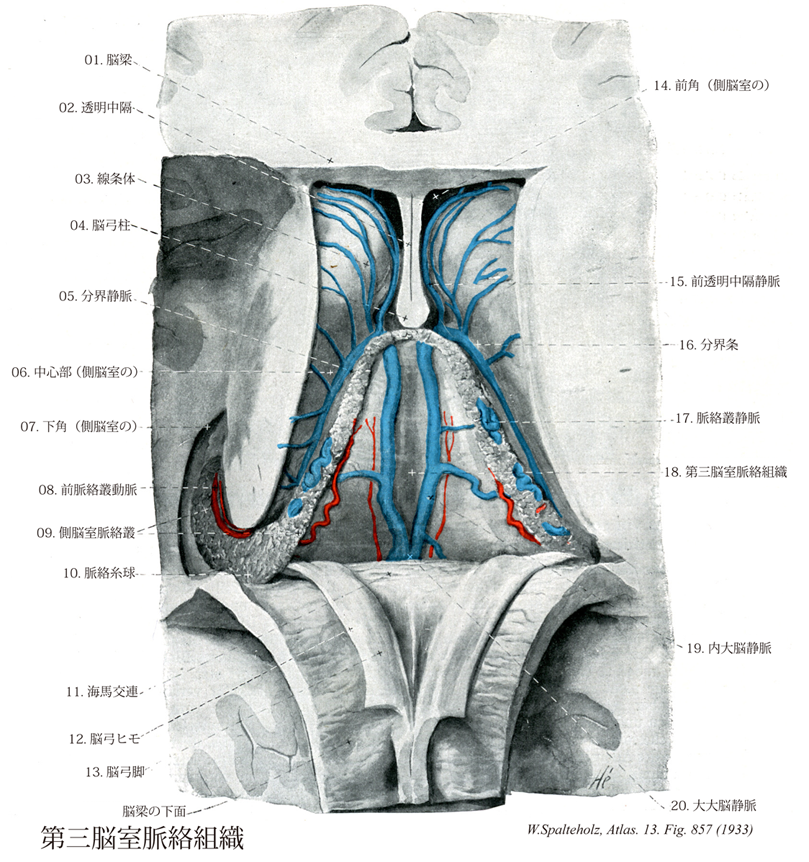
- 857_01【Corpus callosum脳梁 Corpus callosum】 Transverse nerve fibers connecting the two cerebral hemispheres at the base of the longitudinal cerebral fissure.
→(脳梁は左右の大脳皮質、ことに新皮質を結合する線維の集合したもので、系統発生的には最も新しく、ヒトでは非常に発育がよい。その前後経はほぼ7.7cmである。脳梁は正中断では全体としては釣針状で、4つの部分が区別される。後端部は膨大し、脳梁膨大といい、その前方に続いて水平に走る部分を脳梁幹とよぶ脳梁はその前端では強く屈曲し、脳梁膝をつくる。これはさらに後下方にくちばしのように尖って脳梁吻となり、しだいに薄くなって終板に続く。)
- 857_02【Septum pellucidum透明中隔 Septum pellucidum】 Thin dual layer of fibers stretched between the corpus callosum and fornix with an irregular slitlike space between them. It divides the anterior horns of the lateral ventricle from each other.
→(透明中隔は左右の側脳室前角を分離する一対の薄板である透明中隔板と、その間の狭い間隙である透明中隔腔からなる。透明中隔腔は成人ではしばしば閉鎖し、左右の透明中隔板が密着する。透明中隔腔は脳室ではなく、その内面には上皮細胞層が証明されない。透明中隔板は脳梁と脳弓の間に張られているが、発生学的には終脳胞の内側面の一部が脳梁の発達のため前頭葉から分離されたもので、痕跡的な大脳皮質の構造を示す。透明中隔は元来の中隔野の後部の一部で、中隔野には透明中隔のほか、前交連と終板の前にある終板傍回、梁下野、中隔核などが含まれる。)
- 857_03【Corpus striatum線条体[広義の] Corpus striatum】 Structure that is currently viewed as consisting of the putamen. caudate nucleus, pallidum, and fasciculi.
→(線条体は尾状核とレンズ核を意味する。レンズ核はさらに被殻と淡蒼球に区別される。このうち尾状核と被殻は終脳胞の腹外側に出現する神経節丘より同一の細胞群として発生し、その後、のちに発達してくる内包によって二つの部分に隔てられたものである。尾状核と被殻とは内包に横切って走る栓状の灰白質によって互いに連なり、特に前下方では両者は構造的にも同じ細胞構築をもっている。線条体という名称は内包を横切って尾状核と被殻を結んでいる灰白質によってできる縞目と、さらに、尾状核や被殻の中を走る有髄線維の小束によってできる縞目とに基づくものである。したがって、尾状核と被殻とをまとめて線条体Striatumとよび、淡蒼球をPallidumとよんで対比することが多い。解剖学用では、慣用されてきたStriatumという語とは異なる意味内容をもつ語として、Corpus striatumが採用されているが、日本名ではどちらも「線条体」である点は注意を要する。一方、淡蒼球の発生や細胞弧対句はStriatumとは異なる。淡蒼球は有髄神経線維に富むため黄灰白色を呈し、赤みを帯びた暗灰色のStriatumとは肉眼的にも明らかに識別できる。系統発生的視点に立って、尾状核と被殻を新線条体(Neostriatum)、淡蒼球を古線条体(Paleostriatum)、扁桃体を原線条体(Archistriatum)とよぶことがある。)
- 857_04【Column of fornix脳弓柱 Columna fornicis】
→(脳弓柱は没部と出部からでる。没部は左右の乳頭体から始まり、視床下部内を前上方に走り、前交連の後ろで出部に移行する。出部は大脳半球の正中断面で見える部分で、上行するとともにしだいに左右が互いに近づき、ついで後背側方に走り、前方は透明中隔板と癒着している。出部は脳梁幹の下で脳弓体に移行する。)
- 857_05【Superior thalamostriate vein上視床線条体静脈;視床線条体静脈;分界静脈 Vena thalamostriata superior; Vena thalamostriata; Vena terminalis】 It runs in an angle between the thalamus and caudate nucleus, hence its name. It does not receive any branches from the thalamus, but from the rest of the surrounding region. It ends at its union with superior choroid vein in the interventricular foramen.
→(上視床線条体静脈は視床と尾状核の間の溝を前方へ走る長い静脈で、付着板におおわれ、外側から数条の横尾状核静脈が流入し、Monro孔の尾側壁で脈絡叢静脈および透明中隔静脈と合して内大脳静脈となる。)
- 857_06【Central part of lateral ventricle; Body of lateral ventricle中心部;体部;頭頂部(側脳室の) Pars centralis ventriculi lateralis】 Middle portion of the lateral ventricle lying above the thalamus and below the corpus callosum. It contains part of the choroid plexus.
→(側脳室の中心部は中央の最も狭い部分で、Monroの室間孔から側副三角すなわち下角と後角の接合部下に広がる側脳室の体部。上壁は脳梁幹によって作られ、下壁は複雑で、外側から内側方に向かって尾状核尾、分界条、付着板、側脳室脈絡叢、脳弓体などによって形成される。)
- 857_07【Inferior horn of lateral ventricle; Temporal horn of lateral ventricle;下角;側頭角;側頭部(側脳室の) Cornu temporale ventriculi lateralis; Cornu inferius ventriculi lateralis】 It extends with the hippocampus laterally and contains the choroid plexus.
→(側脳室の下角(側頭角)は側頭葉に向かって前下方に突出した部分で、この上外側壁は主として脳梁膨大から放散する脳梁線維からなり、これを壁板という。下壁には側副溝によるたかまり、すなわち側副隆起があり、その後方は三角形をなし、側副三角と呼ばれ、後角まで延びている。内側壁は複雑で、上壁との境界部に尾状核尾および分界条があり、その下に上衣層によっておおわれた側脳室脈絡叢があり、さらにその下には脳弓脚の続きである海馬采がある。)
- 857_08【Anterior choroidal artery前脈絡叢動脈;脈絡叢動脈 Arteria choroidea anterior; Arteria chorioidea】 Artery usually arising from the internal carotid artery. It follows the optic tract posteriorly, enters the choroid plexus at the inferior horn and passes within it to the interventricular foramen. Its tiny branches are usually not visible on angiography.
→(前脈絡叢動脈は後交通動脈の分岐部よりさらに遠位の内頚動脈から通常分岐する。この動脈の特徴は、クモ膜下腔を長い距離を走り、しかも直径が比較的小さいことである。前脈絡叢動脈ははじめ尾方に走って視索を横切り、次いですぐに側頭葉の前内側面に向かって外側方向に走る。つづいて脈絡叢を通過して、側脳室の下角に入る。この動脈が分布する部位として、脈絡叢のほかに、海馬体、淡蒼球の内側および外側領域(すなわち内側部の外がわと外側部の内がわ)、内方後脚の腹側部の大部分、内包のレンズ核後部全体などが含まれる。またこの動脈の小さい枝は、視索、扁桃体の一部、尾状核尾の腹側部、被殻の後部、視床の腹外側領域にも分布する。)
- 857_09【Choroid plexus of lateral ventricle側脳室脈絡叢 Plexus choroideus ventriculi lateralis】 Fringelike, highly vascularized tuft projecting through the choroidal fissure into the lateral ventricle. It extends from the interventricular foramen into the inferior horn.
→(側脳室脈絡叢は脈絡裂から左右の側脳室に突出する血管のふさ。)
- 857_10【Choroid enlargement of lateral ventricle脈絡糸球(側脳室の) Glomus choroideum】 Thickened portion of the choroid plexus in the atrium.
→(側脳室の頭頂部から側頭部への移行部では側脳室脈絡叢の発達が著しいため、とくにこの部分における脈絡叢の集団を脈絡糸球とよぶ。脈絡叢はギリシャの医学者Galenos(129-200頃)がウシやウマの脳を解剖してchoreide plegmata(=chorion-like plaited work)とよんだのに由来する。)
- 857_11【Commissure of fornix脳弓交連;海馬交連 Commissura fornicis; Commissura hippocampi】 Triangular sheet of fibers connecting the crura of fornix below the posterior portion of the corpus callosum. It contains crossing fibers from the fimbriae of hippocampus on both sides.
→(脳弓交連は海馬交連ともよばれる。脳梁後部の下で脳弓脚間にできる三角形の結合野。多数の線維が反対側の脳弓脚へ行き海馬に終わっている。人では発達がわるい。)
- 857_12【Taenia of fornix脳弓ヒモ Taenia fornicis】 Thin, lateral margin of the fornix and attachment site for the choroid plexus of the lateral ventricle.
→(脳弓ヒモは脳弓の側方の薄い縁。側脳室の脈絡叢がつく。)
- 857_13【Crus of fornix脳弓脚 Crus fornicis】 Posterior crus of the fornix that is derived from the fimbria of hippocampus. It encircles the pulvinar and joins with the crus from the opposite side to form the body of fornix.
→(脳弓脚は左右がしだいに離れつつ下外側方に走り、ついで側脳室の下角に向かう。脳梁の下方で左右の脳弓脚の間には脳弓交連がある。)
- 857_14【Frontal horn of lateral ventricle; Anterior horn of lateral ventricle前角;前頭角(側脳室の) Cornu frontale ventriculi lateralis; Cornu anterius ventriculi lateralis】 Portion extending anteriorly from the interventricular foramen. It is bounded medially by the septum pellucidum, laterally by the head of caudate nucleus, superiorly by the body of corpus callosum, and anteriorly and inferiorly by the genu and rostrum of corpus callosum, respectively.
→(側脳室の前角は室間孔より前方の部分、内側壁は透明中隔、外側壁は尾状核頭、前壁及び上下壁は脳梁によってつくられる。)
- 857_15【Anterior vein of septum pellucidum前透明中隔静脈;透明中隔前静脈 Vena anterior septi pellucidi】 It passes from its drainage area, the white matter of the frontal lobe and genu of corpus callosum, through the septum pellucidum to the superior thalamostriate vein.
→(前透明中隔静脈は前頭葉髄質、脳梁膝など流入域から透明中隔をへて視床線条体静脈へ。)
- 857_16【Stria terminalis分界条 Stria terminalis】 Longitudinal band of efferent fibers from the amygdaloid body. It is accompanied by the superior thalamostriate vein in the angle between the thalamus and caudate nucleus.
→(分界条は背側視床と尾状核の間にある狭い白質で、主として扁桃体から起こり、脳弓と平行して走る。この線維の中には視索前野と視床下部と腹内側核でシナプス結合するものがある。)
- 857_17【Choroidal vein脈絡叢静脈 Vena chorioidea】
→()
- 857_18【Tela choroidea of third ventricle第三脳室脈絡組織 Tela choroidea ventriculi tertii】 Thin layer of pia mater covered with ependymal cells extending between the right and left tenia thalami.
→(第三脳室脈絡組織は脳軟膜が大脳半球の後頭葉と小脳の間を通り、さらに脳梁膨大と松果体の間(大脳横裂)から入って第三脳室の上壁を作っているもので、上葉と下葉とからなり、両者の間はクモ膜下組織にによって満たされ、1対の内大脳静脈を含む。第三脳室脈絡組織は全体として頂点を前に向けた三角形をなし、その頂点は脳弓柱、底辺は脳梁膨大、左右の外側辺は付着板の内側縁をなす脈絡ヒモによって作られる。この正中部は第三脳室の上壁を形成し、その下面は第三脳室上衣板でおおわれ、ここから下方に向かい、正中線に沿って左右1対の第三脳室脈絡叢がでる。これらは外側方は視床髄条の表面に視床ヒモをもって付着する。第三脳室脈絡組織の外側部は視床の背側面をおおい、さらに外側方に延びて脳弓(海馬采を含む)と分界条との間にある裂隙、すなわち脈絡裂を通って側脳室の中心部および下角に入り込む。脈絡裂の全長にわたってこの部から側脳室に向かい側脳室脈絡叢が出る。これは内側は脳弓ヒモに、外側は脈絡ヒモに付き、前方は室間孔を通って第三脳室脈絡叢に続き、後方は側脳室の中心部から下角に向かって延びる。中心部と下角の移行部では側脳室脈絡叢が肥厚し、これを脈絡糸球という。)
- 857_19【Internal cerebral veins内大脳静脈;大脳内静脈 Venae internae cerebri; Venae cerebri internae】 Right and left internal cerebral veins. They run in the transverse cerebral fissure, i.e., between the fornix and thalamus or roof of the third ventricle, beginning at the interventricular foramen and ending where they unite from opposite sides to form the great cerebral vein.
→(内大脳静脈は、第三脳室蓋板の脈絡組織内(第三脳室脈絡組織)の正中線近くを走る。この静脈は、室間孔の部位から視床の上内側面を後方に走り、中脳蓋の吻側部のクモ膜下層内で、左右の内大脳静脈が合流して大大脳脳静脈になる。内大脳静脈には、左右それぞれに下記の静脈がはいってくる。①視床線条体静脈(分界静脈)、②脈絡叢静脈、③透明中隔静脈、④視床上部静脈、⑤側脳室静脈である。)
- 857_20Galen, Vein of【Great cerebral vein大大脳静脈 Vena magna cerebri; Vena cerebri magna】 Short vein between the union of the two internal cerebral veins and the beginning of the straight sinus.
→(ガレン大静脈ともよばれる。大大脳静脈は脳梁膨大部の下方に位置し、左右両側の内大脳静脈および脳底静脈が合流してつくられる短い静脈幹である。脳梁膨大部の近くで後方および上方に走行し、大脳鎌と小脳テントの結合部の前方に流し直静脈洞となる。大大脳静脈の全長は平均12mm(範囲8~25mm)と短いが、非常に重要である。大大脳静脈には、1対の脳底内大脳静脈、1対の内大脳静脈、1対の脳底内大脳静脈、1対の脳底静脈、1対の後頭静脈および1対の後脳梁静脈が注ぎ込む。ローマ在住のギリシャの医師Claudius Galen (130-201 ?)による。)