Spalteholz HANDATLAS DER ANATOMIE DES MENSCHEN VON WERNER SPALTEHOLZ
メニューは解剖学(TA)にリンクしてあります。図の番号をクリックすると下記の説明へ、右側の用語をクリックすると解剖学(TA)にジャンプします。
955


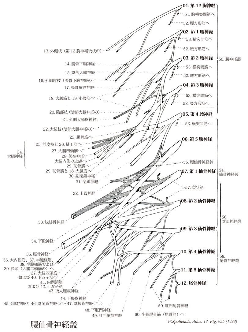
- 955_00【Lumbosacral plexus腰仙骨神経叢 Plexus lumbosacralis】 Communication between the lumbar and sacral plexuses via the lumbosacral trunk.
→(腰仙骨神経叢は第一~第5腰神経の前枝がつくる腰神経叢と腰仙骨神経叢の構成に腰仙骨神経幹(第4腰神経前枝と第5腰神経の前枝の線維の一部)を含むこと、すなわち、腰神経前枝からの線維が構成に加わることは、腰神経叢と仙骨神経叢の間に連続性を作り出す。その結果、2つの神経叢は概念的に腰仙骨神経叢という用語のもとに1つにまとまれられる。)
- 955_01【Twelfth thoracic nerve; T12 spinal nerve; [T12]第12胸神経 Nervus thoracic XII; [T12]】
→()
- 955_01a【Thoracic nerves [T1-T12]胸神経[T1-T12] Nervi thoracici [T1-T12]】 The twelve thoracic spinal nerves that emerge below T1-T12, respectively.
→()
- 955_02【First lumbar nerve; L1 spinal nerve; [L1]第1腰神経 Nervus lumbalis I; [L1]】
→()
- 955_02a【Lumbar nerves [L1-L5]腰神経[L1-L5] Nervi lumbales [L1-L5]】 The five lumbar spinal nerves that emerge below the respective lumbar vertebrae.
→(腰神経は脊髄腰部の両側から対をなして出る5本の神経。)
- 955_03【Second lumbar nerve; L2 spinal nerve; [L2]第2腰神経 Nervus lumbalis II; [L2]】
→()
- 955_04【Third lumbar nerve; L3 spinal nerve; [L3]第3腰神経 Nervus lumbalis III; [L3]】
→()
- 955_05【Fourth lumbar nerve; L4 spinal nerve; [L4]第4腰神経 Nervus lumbalis IV; [L4]】
→()
- 955_06【Fifth lumbar nerve; L5 spinal nerve第5腰神経 Nervus lumbalis V; [L5]】
→()
- 955_07【First sacral nerve; S1 spinal nerve; [S1]第1仙骨神経 Nervus sacralis I; [S1]】
→()
- 955_07a【Sacral nerves and coccygeal nerve [S1-S5, Co]仙骨神経・尾骨神経[S1-S5, Co] Nervi sacralese et nervus coccygeus [S1-S5 Co]】 The five sacral spinal nerves and the one coccygeal spinal nerve.
→(仙骨神経は5対あって、それぞれ、前枝と後枝とに分かれる。はじめの3本は仙骨神経叢に、次の2本は尾骨神経叢に入る。)
- 955_08【Second sacral nerve; S2 spinal nerve; [S2]第2仙骨神経 Nervus sacralis II; [S2]】
→()
- 955_09【Third sacral nerve; S3 spinal nerve; [S3]第3仙骨神経 Nervus sacralis III; [S3]】
→()
- 955_10【Fourth sacral nerve; S4 spinal nerve; [S4]第4仙骨神経 Nervus sacralis IV; [S4]】
→()
- 955_11【Fifth sacral nerve; S5 spinal nerve; [S5]第5仙骨神経 Nervus sacralis V; [S5]】
→()
- 955_12【Coccygeal nervel Coccygeus nerve尾骨神経 Nervus coccygeus】 Last spinal nerve. It emerges between the coccyx and sacrum and communicates with the fourth and fifth sacral spinal nerves.
→(最後の脊髄神経。尾骨と仙骨の間をでて、第四第五仙骨神経と連絡する。 (Feneis))
- 955_13【Lateral branch (of dorsal ramus of 12th thoracic nerve)外側枝(第12胸神経後枝の) Ramus lateralis ramus posterioris (Nervus thoracici XII)】
→()
- 955_14【Iliohypogastric nerve; Iliopubic nerve腸骨下腹神経 Nervus iliohypogastricus; Nervus iliopubicus】 Nerve containing sensory and motor fibers from L1 and T12 that supplies the abdominal muscles. It traverses the psoas major and continues between the transverse abdominal and internal oblique muscles, which it pierces medial to the anterior superior iliac spine.
→(腸骨下腹神経は第12胸神経および第1腰神経よりなり第12肋間神経(肋下神経)の下方を、これと平行に前下方に走り、前腹筋に、皮枝を下腹部と臀部との皮膚に与える。大腰筋を斜めに貫き、腹横筋および内腹斜筋の間にでて、上前腸骨棘の内側でこれらの腹筋を貫く。)
- 955_15【Genitofemoral nerve陰部大腿神経 Nervus genitofemoralis】 Nerve arising from L1 and L2 that pierces and lies on top of the psoas major.
→(陰部大腿神経は第12胸神経、第1および第2腰神経よりなる細い神経で、大腰筋の中で2枝に分かれ、一本は精索(子宮円索)に沿って陰嚢(大陰唇)に分布し、他の一本は外腸骨動脈に沿って下行枝し、鼡径靱帯の下を通り、伏在裂孔から出て大腿上部の内側部皮膚に分布する。陰部大腿神経は精巣挙筋を支配しいている。神経が分布する大腿近位部の内側部皮膚を擦ると挙睾筋(精巣挙筋)の反射的収縮によって精巣は上昇する(挙筋反射cremasteric reflex)。)
- 955_16【Lateral cutaneous branch of iliohypogastric nerve外側皮枝(腸骨下腹神経の) Ramus cutaneus lateralis (Nervus iliohypogastrici)】 Branch that can extend as far as the lateral gluteal region.
→(腸骨下腹神経の外側皮枝は臀部外側まで達することある。)
- 955_17【Ilioinguinal nerve腸骨鼡径神経 Nervus ilioinguinalis】 It usually arises from L1 and emerges at the lateral border of the psoas. It travels between the kidney and quadratus lumborum, then between the transverse abdominal and internal oblique muscles, continuing through the inguinal canal.
→(腸骨鼡径神経は腸骨下腹神経の下方を、これと平行に走り、筋枝を側副筋に与えたのち精索(女性では子宮円索)とともに、鼡径管を通って鼡径部の皮下に現れ、陰嚢(女性では大陰唇)に分布する(前陰嚢神経または前陰唇神経)。)
- 955_18【Psoas major muscle大腰筋 Musculus psoas major】 o: Lateral aspect of vertebral bodies T12 and L1-L4, costal process of L1-L5. I: Lumbar plexus.
→(横隔膜の内側弓状靱帯の後を走る大腰筋は浅深の2頭を持つ。浅頭は第12胸椎~第4腰椎の椎体と椎間円板から起こり、深頭は全腰椎の肋骨突起から起こる。これら2頭の間には腰神経叢の枝が何本も走っている。大腰筋は下方では腸骨筋と共に鼡径靱帯の後をくぐって腸腰筋の一部となって大腿に下る。)
- 955_19【Psoas minor; Psoas minor muscle小腰筋 Musculus psoas minor】 o:Vertebral bodies of T12 and LI. i: Fascia iliaca. I: Lumbar plexus.
→(大腰筋の前面には、小腰筋が40~50%の頻度で存在する。この細長い筋は、上位腰椎の椎体から起こり、その腱は腸骨筋の表面の筋膜(腸骨筋間)に放散しながら恥骨櫛に終わる(すなわち小腰筋は骨盤の外には出ない)。第3の寛骨内筋、その起始は第12胸椎と第1腰椎である。長い腱は大腰筋上を尾側へ向かい、腸腰筋膜や、特に腸恥骨弓に放散する。小腰筋は約半例で欠損する。人種差は認められないが、概して女性に欠ける率が多い。また小腰筋は重複することがある。)
- 955_20【Genital branch of genitofemoral nerve陰部枝(陰部大腿神経の);外精神経 Ramus genitalis (Nervus genitofemoralis); Nervus spermaticus externus】 Branch running through the inguinal canal that supplies the cremaster, scrotal skin (labium majus), and adjacent skin of the thigh.
→(陰部大腿神経の陰部枝は深鼡径輪を通って陰嚢前面、大陰唇、隣接する大腿の皮膚に分布し、運動枝を精巣挙筋に送る。)
- 955_21【Lateral cutaneous nerve of thigh; Lateral femoral cutaneous nerve; Fibular femoral cutaneous nerve外側大腿皮神経;大腿の外側皮神経;腓側大腿皮神経 Nervus cutaneus femoris lateralis; Nervus cutaneus femoris fibulalis】 It arises from L2 and L3 and emerges at the lateral border of the psoas. It travels beneath the iliac fascia through the lateral part of the muscular space and either deep or superficial to the sartorius, continuing to the skin of the lateral aspect of the thigh.
→(外側大腿皮神経(L2,L3)は、大腰筋の外側縁から出現し、壁側腹膜と腸骨筋膜(この神経の支配を受ける)の後方で腸骨筋を斜めに越える。上前腸骨棘の方向へ進み、鼡径靭帯外側端を越えるるか、あるいはこれを貫通して大腿に入る。ついで、外側大腿皮神経は、縫工筋近位部を越えるか、あるいは貫通した後、大腿筋膜の深部を下行する。鼡径靱帯の下方約10cmで大腿筋膜を貫くが、それ以前に、この神経を覆う皮膚に数多くの小枝を出す。外側大腿皮神経の終末枝は大腿骨大転子と膝との間で、大腿の前外側面を覆う皮膚と筋膜に分布する。)
- 955_22【Femoral branch of genitofemoral nerve大腿枝(陰部大腿神経の);腰鼡径神経 Ramus femoralis (Nervus genitofemoralis); Nervus lumboinguinalis】 Branch passing through the vascular space and continuing through the saphenous opening to supply the overlying skin.
→(陰部大腿神経の大腿枝は血管裂孔(大体動脈と腸恥筋膜弓の間)を通り、伏在裂孔をへて、大腿前面最上部の皮膚へ分布する。)
- 955_23【Iliacus muscle腸骨筋 Musculus iliacus】 o: Iliac fossa, hip joint capsule. I: Femoral nerve, lumbar plexus.
→(腸骨稜の下方に腸骨筋膜に囲まれて存在する。腸骨窩に起始し、大腰筋との共同腱によって小転子前面と股関節嚢に停止する。腰神経叢によって神経支配され、大腿の屈曲の内旋に作用する。)
- 955_24【Femoral nerve大腿神経 Nervus femoralis】 Thickest branch of the lumbar plexus, which arises from L2-L4. It emerges at the lateral border of the psoas and travels in the muscular space between it and the iliacus. It divides below the inguinal ligament.
→((Netter)大腿神経(L2,L3,L4)は、腰神経叢から出る最大の枝で、腸骨筋と大腿前面の筋群を支配し、股関節や膝関節および周囲の血管へ枝を送り、また下肢の前内側面に皮枝を出す。大腿神経は、第2~4腰神経前枝の背側部から起こり(図10)、大腰筋を下外方へ貫通し、ついで大腰筋と腸骨筋間の溝を走行しながらこれらの筋に支配枝を出す。大腿神経は、鼡径靱帯の後方を通り、大腿部に入る。大腿三角では大腿動脈鞘の外側に位置し、ここで筋枝と皮枝に分かれる。筋枝は、恥骨筋、縫工筋および大腿四頭筋を支配する。恥骨筋へ行く筋枝は、鼡径靱帯の高さで起こる。縫工筋への筋枝は、この筋の上部2/3に入る。また数本の枝は、大腿神経前皮枝と起始を同じくしている。大腿四頭筋への支配枝は、図のごとく支配する。すわなち大腿直筋と外側広筋への枝は、両筋の後面へ入る。また、中間広筋への枝は、その前面から中間広筋に入り、中間広筋を貫いて下部にある膝関節筋を支配する。さらに、内側広筋への枝は、内転筋管を大腿動静脈と伏在神経の外側に沿って様々な長さを走りながら、次々に内側広筋へ枝を出し、そのうちの何本かは中間広筋や膝関節筋に終わる。 大腿神経の前皮枝は、大腿三角に起こる。鼡径靱帯より8~10cm遠位で、この神経のすべての枝は大腿筋膜を貫き、膝関節の高さまで下行する。走行の間に、大腿前面および内側面を覆う皮膚と筋膜に枝を送る。伏在神経は、大腿神経の最大かつ最長の枝であり、大腿三角の高さで起こり、大腿動静脈の外側に沿って大腿三角内を下行し、内転筋管に入る。ここでこの神経は大腿動静脈を斜めに越え、大内転筋下端の前面で、これらの動静脈内側に位置するに至る。内転筋管内において、伏在神経は、大腿神経前皮枝および閉鎖神経と交通して縫工筋下神経叢を形成する。内転筋管下端では、この神経は、大腿動静脈から離れて膝蓋下枝を生じる。この枝は、縫工筋の後縁を回り、大腿筋膜を貫いて走行を続け、膝関節や膝蓋靱帯の内面および前面を覆う皮膚に分布する。膝蓋下枝は大腿神経前皮枝および外側大腿皮神経より枝を受けて膝蓋神経叢を作る。伏在神経は、膝関節の内側面を下行し、縫工筋と薄筋の間で大腿筋膜を貫く。この後、大伏在静脈の近傍で下腿の内側面を下行し、内側下腿皮枝を出す。伏在神経は、下腿の下部でさらに2枝に分かれる。小さな枝は脛骨内側縁に沿って足首まで下げる。他方、大きい枝は、内果前面を越え、足の内側面と足背を覆う皮膚と筋膜に分布する。関節枝は、大腿直筋の支配枝から起こり、外側大腿回旋動脈の関節枝と共に股関節に至る。大腿の広筋群への支配枝からの小枝および伏在神経からの小枝が、膝関節を支配する。)
- 955_25【Anterior cutaneous branches of femoral nerve前皮枝(大腿神経の) Rami cutanei anteriores (Nervus femoralis)】 Cutaneous nerves supplying the distal three-fourths of the anterior side of the thigh as far as the patella.
→(大腿神経の前皮枝は大腿の前面と内側面に分枝し、大腿前面を膝蓋にいたる下部3/4の皮膚へ分布し、感覚を伝える。)
- 955_26【Sartorius muscle縫工筋 Musculus sartorius】 o: Anterior superior iliac spine, i: Medial to the tibial tuberosity. Flexion, abduction, lateral rotation at the hip joint; flexion and medial rotation at the knee joint. I: Femoral nerve.
→(縫工筋は大腿前部浅層の筋。上前腸骨棘からラセン状に大腿前面と内側面を走る。大腿筋膜がつくる筋膜の鞘に包まれている。その弓状走行により、同筋は背側にある膝関節の屈曲軸を横切ることになる。停止腱は遠位かつ腹側へ斜走し、脛骨内側面(脛骨粗面のうしろ)が鵞足に、また下腿筋膜に停止する。鵞足は縫工筋(浅層)、薄筋および半腱様筋(深層)の末広がりの停止腱が1カ所に集まってできる。その様子は鵞の足の水掻きが折れ重なったようにみえる。鵞足は内側側副靱帯とは鵞足包で隔てられ、その腱線維は脛骨内側面に放散する。また、浅層の線維は下腿筋膜に続く。かつて縫工(仕立屋)は作業をするときに、あぐらをかくように脚をむく姿勢をとった。このように、大腿を屈曲・外転・外旋し、かつ膝を屈曲するのに、縫工筋が働くと考え、名付けられた。)
- 955_27【Quadriceps femoris muscle大腿四頭筋 Musculus quadriceps femoris】 Muscle group composed of the following four muscles. Its tendon extends to the patella and continues as the patellar ligament to the tibial tuberosity. Extension at the knee joint. I: Femoral nerve.
→(大腿四頭筋は大腿前部の筋で下腿を伸展するほとんど唯一の筋であって、膝蓋骨と膝蓋靱帯を介して脛骨粗面につく。大腿四頭筋は4頭を持つ。大腿直筋は股関節と膝関節をまたぎ、これに大腿骨のほとんどを包む3つの広筋が合流する。4つの筋は広い共同腱を作り、その中に膝蓋骨が種子骨として埋まっている。膝蓋骨尖からは膝蓋靱帯が停止腱の続きとしてあり、脛骨粗面に達する。膝蓋骨によって大腿四頭筋腱は膝関節屈曲軸から遠くなり、その分、筋の回転力が増加する。膝蓋支帯は大腿四頭筋の側方腱で、縦長の紐状物として膝蓋骨側方を走り、脛骨上縁に付く。膝蓋骨が大腿骨膝蓋面から滑脱するのを防ぎ、大腿四頭筋の伸展作用の一部を脛骨に伝える。)
- 955_28【Saphenous nerve伏在神経 Nervus saphenus】 Longest, purely sensory branch of the femoral nerve. It begins in the femoral triangle, passes beneath the 「vastoadductor membrane,」 which it pierces, continues between the sartorius and gracilis to beneath the skin, and then travels with the great saphenous vein as far as the medial side of the foot.
→(大腿三角から足に至る大腿神経の枝。伏在神経は大腿動脈の外側を沿って走り、動脈とともに内転筋管内を下降する。膝関節の内側で皮下にでて大伏在静脈に沿って下行し、下腿と足背との内側面の皮膚に分布する。)
- 955_29【Pectineus muscle恥骨筋 Musculus pectineus】 o: Pectineal line of pubis. i: Pectineal line of femur, linea aspera. Flexion, adduction, and medial rotation at the hip. I: Femoral and obturator nerves.
→(恥骨筋は腸恥隆起-恥骨結節間の恥骨上肢から起こり、大腿骨の恥骨筋線に停止する。この筋はもとももと腸腰筋群と同じ原基に由来する。本筋の構成に内転筋群がどの程度関わるかには個体差がある。恥骨筋は腸骨筋膜の延長部分である恥骨筋膜におおわれ、腸腰筋とともに腸恥窩には大腿動静脈が通る。)
- 955_30【Accessory obturator nerve副閉鎖神経 Nervus obturatorius accessorius】 Occasional accessory nerve arising from L3 and L4 that supplies the pectineus and hip joint.
→(副閉鎖神経(L3,L4)は、必ずしも存在しない。存在する場合には、この神経は小さく、第3、第4腰神経の前枝腹側部より生じる。副閉鎖神経は、大腰筋の内側縁に沿って下行し、恥骨上枝を越えて恥骨筋の後方に至る。この神経は、恥骨筋の神経支配を補佐して終わるが、股関節への支配枝や閉鎖神経前分枝に加わる枝を派生することがある。)
- 955_31【Obturator nerve閉鎖神経 Nervus obturatorius】 It arises from L2-L4 and travels beneath the psoas, posterior to the internal iliac artery, lateral to the ureter, continuing through the obturator canal to the adductors and the skin of the medial aspect of the thigh.
→(閉鎖神経は第二・第三・第四腰神経からなる腰神経叢から起こり、垂直に下行し、大腿筋の内側縁から出て、総腸骨動脈の後側を通って骨盤孔に入る。骨盤の側壁内面に沿って走り、閉鎖動静脈とともに閉鎖管を抜けて大腿上部の内側部に出る。前枝は長内転筋と薄筋の間に現れ、大腿皮膚の下3分の2へ分布。 筋枝は大腿の内転筋に分布する。大腿の内側部皮膚と内転筋分に分布する。皮枝は大腿内側の皮膚に分布する。関節枝は股関節と膝関節とに分布する。)
- 955_32【Superior gluteal nerve上殿神経;上臀神経 Nervus gluteus superior】 Nerve arising from L451 that passes through the greater sciatic foramen cranial to the piriformis and then between the gluteus medius and minimus to the tensor muscle of fascia lata, which it supplies as well as the above-mentioned muscles.
→(上臀神経はL4~S1より起こる。大坐骨孔を梨状筋の上で通り(梨状上孔)臀部に入り中および小臀筋の間を抜け大腿筋膜長筋へいたる。これらの筋肉を支配する。)
- 955_33【Common fibular nerve; Common peroneal nerve総腓骨神経 Nervus fibularis communis; Nervus peroneus communis】 Branch of the sciatic nerve arising from L4-S2. It accompanies the tendon of the biceps femoris to posterior to the head of fibula and then crosses obliquely forward between the skin and the fibula.
→(総腓骨神経はL4~S2より起こる。坐骨神経の2終枝のうちの細い方である。大腿の下1/3の高さから始まり、膝窩内を下腿への皮神経を出しながら、大腿二頭筋の停止腱の後内側縁に沿って腓骨頭の後面に達し、ここで下腿筋の下層にもぐり込む。腓腹筋外側頭よりも浅層でこれを横切り、膝窩を離れる。以上の経過中に総腓骨神経は腓骨頚外側面で皮膚のみに被われた状態となるため、体表よりこの神経を容易に触知できる。そののち総腓骨神経は腓骨頭の後方をやや下行してから急に向きを変えて長腓骨筋内に侵入し、そこで浅腓骨神経および深腓骨神経に分かれる。)
- 955_34【Inferior gluteal nerve下殿神経;下臀神経 Nervus gluteus inferior】 Nerve arising from L5-S2 that travels through the infrapiriform foramen and supplies the gluteus maximus.
→(下臀神経はL5~S2より起こる。大坐骨孔を通り梨状筋下部(梨状下孔)から臀部に入る。後大腿神経に近接した走行を示したのちに、下臀神経は大臀筋を支配する。)
- 955_35【Tibial nerve脛骨神経 Nervus tibialis】 Second terminal branch of the sciatic nerve arising from L4-S3. It travels through the popliteal fossa, passes deep to the tendinous arch of the soleus, and accompanies the posterior tibial artery around the medial malleolus to the sole of the foot.
→(脛骨神経はL4~S3より起こる。坐骨神経の第二の終枝。膝窩を通りヒラメ筋腱弓の下をすぎ後脛骨筋とともに内果をまわり、足底へ達す。下腿の屈筋群、足底の諸筋、下腿の後面と足底の皮膚に分布するが、次の神経はいずれも脛骨神経の末梢枝である。①下腿骨間神経(下腿骨間膜の後縁に沿って走り、足関節のあたりに達する)、②内側被覆皮神経、腓腹神経、外側足背神経(ひとつづきのもので下腿後面から足背外側部の皮膚に分布)、③内側足底神経と外側足底神経(ともに足底の諸筋に分布する枝を出したあと、趾の足底面や足底の皮膚に分布するため、総底側趾神経に枝分かれし、固有底側趾神経となっておわる)。)
- 955_36【Adductor magnus muscle大内転筋 Musculus adductor magnus】 o: Inferior pubic ramus, ramus of ischium. i: Medial lip of linea aspera and, via a long tendon, the medial epicondyle. Adduction, lateral rotation, and extension at the hip joint. I: Obturator and tibial nerves.
→(大内転筋は内転筋群の中で最強、人体中最大の筋の1つである。他の内転筋の背側、恥骨下枝および隣接する坐骨枝から坐骨結節までを起始とする。他の起始をもつ部分はほとんどが粗線内側唇に筋性停止する。)
- 955_37【Semitendinosus muscle半腱様筋 Musculus semitendinosus】 o: Ischial tuberosity. i: Medial surface of tibia. Extension at the hip joint. Flexion and medial rotation at the knee joint. I: Tibial nerve.
→(半腱様筋は大腿二頭筋長頭の起始近くの坐骨結節から起こり、鵞足を介して脛骨近位端内側面および下腿筋膜に終わる。半腱様筋は半膜様筋によってつくられた溝の中を遠位へ向かう。長い停止腱は大腿部ですでに始まり(ここから“半腱様”の名がつけられた)、鵞足の深層へと放散する。)
- 955_38【Semimembranosus muscle半膜様筋 Musculus semimembranosus】 o: Ischial tuberosity. i: Medial condyle of tibia and oblique popliteal ligament. It is partly covered by the semitendinosus muscle. Extension at the hip joint; flexion and medial rotation at the knee joint. Tenses the knee joint capsule. I: Tibial nerve.
→(半膜様筋は大腿二頭筋長頭と大腿方形筋の起始の間の坐骨結節から起こる。脛骨内側顆、膝関節包後壁および膝窩筋の筋膜に停止する。半膜様筋は中4分の2のみが筋性である。起始腱は広い腱性の板をなし、停止腱も同じ平板である。3本の腱様の索として終わる。脛骨への索は腹側で迂回し、内側側副靱帯の下の脛骨内側顆に付く。中央の索は筋の方向を受け継ぎ、一部は脛骨近位端後面に、一部は膝窩筋の筋膜に付く。腓骨への索は膝関節包の後壁を補強し、斜膝窩靱帯として大腿骨外側顆に向かって外側へ射創する滑液包が通常同筋の停止腱と脛骨内側顆の間にある。)
- 955_39【Long head of biceps femoris muscle長頭(大腿二頭筋の) Caput longum (Musculus biceps femoris)】 o: Ischial tuberosity. Extension at the hip joint. I: Tibial nerve.
→(大腿二頭筋の長頭は半腱様筋とともに坐骨結節後面から起こる。短頭は粗線外側唇の中1/3から起こる。同レベルの粗線内側唇に停止する長内転筋の線維方向を短頭は引き継ぐ。両頭の共通停止腱は腓骨頭に止まる。腱線維の一部は脛骨外側顆および下腿筋膜に達する。大腿二頭筋腱は腱下包によって外側側副靱帯と隔てられる。)
- 955_40【Gemellus inferior muscle; Inferior gemellus muscle下双子筋;結節双子筋 Musculus gemellus inferior; Musculus gemellus tuberalis】 o: Ischial tuberosity. i: Tendon of obturator internus, trochanteric fossa. Lateral rotation, adduction, abduction. I: Sacral plexus.
→(この小さな下双子筋は、坐骨結節上縁により起こり内閉鎖筋腱に停止しする。仙骨神経叢からの大腿方形筋への直接枝による神経支配を受け、大腿骨を外旋させる作用を示す。)
- 955_41【Obturator internus muscle; Internal obturator muscle内閉鎖筋 Musculus obturator internus】 o: Internal surface of obturator membrane and surrounding area, i: Trochanteric fossa of greater trochanter. Lateral rotation, abduction, adduction. I: Sacral plexus.
→(内閉鎖筋と2つの双子筋は発生的にはひとまとまりである。内閉鎖筋はその起始を骨盤腔内へ移し、閉鎖膜上および閉鎖孔の骨性枠から起こるに至った。小坐骨孔縁(軟骨でおおわれている)が視点となり、内閉鎖筋包が介在し、ここで急に走行を骨盤外へ変える。骨盤の外にある部分は3分筋のうちの2頭、つまり上下の双子筋を多少なりともおおう。上双子筋は坐骨棘を発し、下双子筋は坐骨結節を発する。内閉鎖筋の停止腱の上下縁にはそれぞれ上下の双子筋が合流し、転子窩に終わる。骨盤内にある内閉鎖筋は強い内閉鎖筋膜に包まれ、これが肛門挙筋の起始となる。内閉鎖筋膜は肛門挙筋起始部では弓状をした腱様の筋膜束(肛門挙筋腱弓)で補強さえている。肛門挙筋腱弓よりも上で、内閉鎖筋は小骨盤の筋性壁をつくり、その筋膜は壁側骨盤筋膜の一部となる。これより下では内閉鎖筋とその筋膜は外側部において、骨盤底の下にある結合組織性の部位、すなわち坐骨直腸窩を区画する。)
- 955_42【Gemellus superior muscle; Superior gemellus muscle上双子筋;棘双子筋 Musculus gemellus superior; Musculus gemellus spinalis】 o: Ischial spine, i: Tendon of obturator internus muscle and trochanteric fossa. Lateral rotation, adduction, abduction. I: Sacral plexus.
→(この小さな上双子筋は坐骨棘より起こり内閉鎖筋腱に停止する。仙骨神経叢からの、内閉鎖筋へ直接枝による神経支配を受け、大腿骨外旋作用を示す。)
- 955_43【Posterior cutaneous nerve of thigh; Posterior femoral cutaneous nerve後大腿皮神経;後皮神経(大腿の) Nervus cutaneus femoris posterior】 Nerve arising from SI-S3 that travels through the greater sciatic foramen distal to the piriformis and supplies the skin on the posterior side of the thigh as well as the proximal part of the leg.
→(後大腿皮神経は仙骨神経叢のS1~S3より起こる。大坐骨孔を通り梨状筋の下で臀部にあらわれる。この神経は坐骨神経と大臀筋ではさまれながら臀部を下行し、大腿二頭筋よりも浅層を走りながらこれを横切り大腿背面の深筋膜内を走る。膝窩に達してから後大腿皮神経本幹は深筋膜を貫き皮膚に向かう。)
- 955_44【Inferior clunial nerves; Inferior cluneial nerves下殿皮神経;下臀皮神経 Nervi clunium inferiores】 Cutaneous branches ascending along the inferior margin of the gluteus maximus.
→(下臀皮神経は大臀筋の下縁を上行する皮枝。(Feneis))
- 955_45【Perineal nerves会陰神経 Nervi perineales】 Collective term for the following two perineal nerves.
→(会陰神経は球海綿体筋、坐骨海綿体筋、浅会陰横筋およびこれらの筋をおおう皮膚に分布したあと、陰嚢または陰唇の後部に分布する後陰嚢神経または後陰唇神経となる。)
- 955_46【Dorsal nerve of penis♂陰茎背神経(♂) Nervus dorsalis penis♂】 Paired nerve lying on the dorsum of penis that also sends branches to its inferior aspect.
→(陰茎背神経は深会陰横筋に分枝したあと、これを貫いて陰茎または陰核背面に達し、亀頭、包皮、尿道粘膜などに分布する。)
- 955_47【Dorsal nerve of clitoris♀陰核背神経(♀) Nervus dorsalis clitoris♀】 Small nerve that corresponds to the dorsal nerve of the penis.
→(陰核背神経は男性の陰茎背神経に相同な小神経で尿生殖隔膜に分枝したあと、これを貫いて陰核背面にに達し、陰核亀頭、包皮および尿道粘膜などに分布する。)
- 955_48【Inferior anal nerves; Inferior rectal nerves下肛門神経;下直腸神経;肛門神経 Nervi anales inferiores; Nervi rectales inferiores; Nervi anales】 Fibers arising from the third and fourth sacral spinal nerves that supply the external anal sphincter and anal skin.
→(下直腸神経は第三および第四仙骨神経よりでる線維で、これは坐骨直腸窩を内側に横切る神経であり、同名動静脈とともに外肛門括約筋、肛門管下半の粘膜、会陰皮膚などに分布する。)
- 955_49【Nerve to levator ani muscle of sacral plexus; Branch to levator ani muscle肛門挙筋神経;肛門挙筋への枝(仙骨神経叢の) Nervus musculi levatoris ani】
→()
- 955_49a【Levator ani muscle肛門挙筋 Musculus levator ani】 Principle muscle of the pelvic diaphragm. It is derived from the abdominal wall musculature and permeated by smooth-muscle cells. I: Sacral plexus, S2-S5. It consists of the following parts.
→(肛門挙筋の丈夫な前部(恥骨尾骨筋)は分界線直下の恥骨の内面から起こり、薄い後部(腸骨尾骨筋)は腸骨から起こる。その起始腱は内閉鎖筋筋膜に接して移行し、閉鎖筋膜から発する腱束を受ける。これらの線維の起始部では腱性の係留物(肛門挙筋腱弓)により強化されている。左右両側で恥骨尾骨筋の内側線維束は挙筋脚を形成している。それらの線維束は背方と尾方、また直腸の前では外側を通り、それぞれ会陰の中心腱へ放散する薄い前直腸線維束や前立腺挙筋として前立腺筋膜(あるいは恥骨腟筋として腟壁)へと分かれる。それより鼻側にある肛門挙筋の線維束は恥骨直腸筋として直腸の背側を取り囲み、反対側の線維と共にループを形成する。恥骨尾骨筋の外側束は尾骨と仙骨の背側に広がる。腸骨尾骨筋の筋線維は尾骨と仙骨に付き、また肛門と尾骨の間では強靱な線維束である肛門尾骨靱帯に付いている。)
- 955_50【Lumbar plexus; Lumbar nerve plexus腰神経叢 Plexus lumbalis】 Plexus formed by the anterior rami of L1-L3 and portions of T12 and L4. Its nerves lie mainly along the inferior abdominal wall and anterior surface of the leg.
→(腰神経叢は第12胸神経、第1~第3腰神経、第4腰神経の一部の神経線維により、大腰筋の後方と筋内で形成される。この神経叢の神経は下肢の前面に至る。)
- 955_51【Thoracic intertransversarii; Thoracic intertransversarii muscles胸横突間筋;胸後横突間筋 Musculi intertransversarii thoracis; Musculi intertransversarii posteriores thoracis】 Usually absent.
→(胸横突間筋は深背筋上部の筋の一つ。起始と停止は、外側部は頚椎横突起の後結節、内側部は胸椎の横突起にはじまり、一つ上位の胸椎の横突起につく。外側部は胸神経の前枝、内側部は胸神経の後枝からの神経支配を受ける。作用として胸椎の外転。)
- 955_52【Quadratus lumborum muscle腰方形筋 Musculus quadratus lumborum】 o: Iliac crest, i: Twelfth rib, costal processes of lumbar vertebrae L1-L4. Draws ribs inferiorly, lateral flexion. I: Intercostal nerve of twelfth rib, lumbar plexus.
→(腰方形筋の起始は腸腰靱帯、腸骨稜の後部。停止は最後の肋骨の下縁、上位4個の腰椎横突起。機能として一番下の肋骨の下制(ひきさげ)、体幹屈曲の補助、一側単独で働くときは脊柱の側屈、呼吸運動の関与は疑問。神経支配は肋下神経、第1,2,3腰神経。動脈は腸腰動脈の腰枝から受ける。)
- 955_53【Intertransversarii muscles横突間筋;横突間筋群 Musculi intertransversarii dorsi proprii】 Muscles that connect the transverse processes of consecutive vertebrae. I: Posterior rami of spinal nerves of C1-C6; L1-L4.
→(横突間筋の起始と停止は隣り合う脊椎横突起の間にわたっている細い筋肉束、頚椎と腰椎に最もはっきりみられる。機能として恐らく脊柱の側方屈曲の補助。神経支配は脊髄神経の後枝。腰椎および下位胸椎部の外側横突間筋と頚椎部の前後横突間筋は例外であって脊髄神経前枝の支配を受けている。動脈は肋頚動脈の深頚枝、後肋間動脈と腰動脈の筋枝から受ける。固有背筋(脊髄神経後枝の支配)のほかに、体幹腹外側筋(脊髄神経前枝の支配)に属するものも含まれる。横突間筋のうち固有背筋に属するものは頚部、腰部、胸部の上下端部にある:頚後横突間筋の内側部(後結節の間)、胸横突間筋、腰内側横突間筋(乳頭突起や副突起の間)。体幹腹外側筋に属するものは頚部と腰部にある:頚後横突間筋の外側部(後結節の間)、頚前横突間筋(前結節の間で脊髄神経前枝の前)、腰外側横突間筋(肋骨突起の間にあってかなり発達する)。)
- 955_54【Sacral plexus; Sacral nerve plexus仙骨神経叢;坐骨神経叢 Plexus sacralis; Plexus ischiadicus】 Plexus formed by L5-S3 and part of L4 and S4. It lies on the anterior aspect of the piriformis deep to the fascia and sends nerves to the posterior aspect of the leg.
→(仙骨神経叢は第4腰神経、第5腰神経、第1~第5仙骨神経の前枝がつくる脊髄神経叢であり、交通枝で交感神経と連絡する。仙骨神経叢の神経は、次の枝を出す。①梨状筋、内閉鎖筋、双子筋、大腿方形筋への筋枝、②中臀筋、小臀筋、大腿筋膜張筋に分布する上臀神経、③大臀筋に分布する下臀神経、④下臀部、会陰部、および大腿後面の皮膚に分布する後大腿皮神経、⑤坐骨神経。第2仙骨神経から第4仙骨神経で形成される神経叢を陰部神経叢pudendal trunkとし、それより上方の神経叢を坐骨神経叢sciatic trunkということもある。仙骨神経から起こる神経は臀部・下肢に分布する神経と骨盤部に分布する神経とに大別できる。)
- 955_55【Lumbosacral trunk腰仙骨神経幹 Truncus lumbosacralis】 Communication with the lumbar plexus formed by L5 and part of L4.
→()
- 955_56【Pudendal trunk陰部神経叢 Plexus pudendalis】
→(第二~第四仙骨神経の前枝が構成する神経叢をこのようによぶことがある。骨盤内蔵(内臓枝)と会陰部(陰部神経)とを支配する。)
- 955_57【Piriformis muscle梨状筋 Musculus piriformis】 o: Anterior surface of sacrum, i: Greater trochanter, medial aspect of the apex. Abduction, extension, and lateral rotation at the hip joint. 1: Sacral plexus.
→(梨状筋は骨盤の後側の深層にある回旋筋で仙骨の骨盤筋膜(前仙骨孔およびそれらの外側の)と仙腸関節の関節包から、大坐骨切痕上縁に由来する線維束とともに、起始する。大坐骨孔を通過して、同孔を梨状筋上孔と下孔に分け、大腿筋の深層を骨盤外側をおおって走り、大転子先端の内側に至る。広い起始に始まり、梨状筋は次第に収束して細い停止腱となる。股関節と停止腱は滑液包により隔てられる。坐骨神経の梨状筋枝から支配を受け、股関節の外転、伸展および外旋を行う。)
- 955_58【Coccygeal plexus尾骨神経叢 Plexus coccygeus】 Nerve plexus formed by S5, part of S4, and the coccygeal nerve. It supplies the skin overlying the coccyx.
→(尾骨神経叢は第五仙骨神経および尾骨神経からなる小神経叢。肛門尾骨神経が起こる。 (Netter)第4,第5仙骨神経と尾骨神経の各前枝下部が結合して、小さな尾骨神経叢を作る。尾骨神経叢は、尾骨筋と肛門挙筋への筋枝および肛門と尾骨間を覆う皮膚に分布する細い肛門尾骨神経が派生する。)
- 955_59【Anococcygeal nerves肛門尾骨神経;肛尾神経 Nervus anococcygeus】 Several thin nerves from the coccygeal plexus that penetrate the anococcygeal ligament and supply the overlying skin.
→(尾骨神経叢よりでる多数の小枝。肛門尾骨靱帯を貫き、その上の皮膚へ分布。 (Feneis))
- 955_60【Ischiococcygeus muscle; Coccygeus muscle; Coccygeal muscle坐骨尾骨筋;尾骨筋 Musculus ischiococcygeus; Musculus coccygeus】 Muscle fibers that fan out from the ischial spine to the lateral surfaces of the sacrum and coccyx. They are joined with the sacrospinal ligament.
→(仙骨下端と尾骨を結ぶ筋で、ヒトでは退化的に(坐骨)棘尾骨筋に相当し、その浅(外)部が仙棘靱帯になり、深(内)部が尾骨筋に変化し、肛門挙筋とともに尾骨隔膜を形成する。前後の仙尾筋は、それぞれ腹側と背側の尾筋に相当する。後仙尾筋は固有背筋の最下部である。)