Spalteholz HANDATLAS DER ANATOMIE DES MENSCHEN VON WERNER SPALTEHOLZ
メニューは解剖学(TA)にリンクしてあります。図の番号をクリックすると下記の説明へ、右側の用語をクリックすると解剖学(TA)にジャンプします。
972


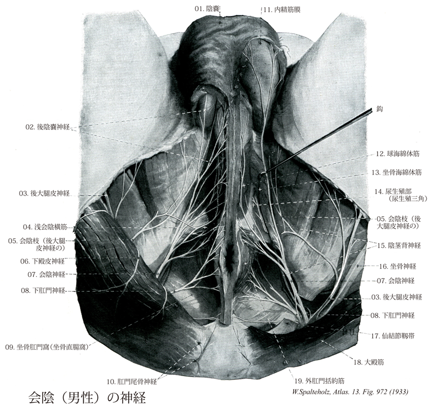
- 972_00【Perineum会陰 Perineum】 A term that has different uses: Soft-tissue bridge between the anus and genitalia. In topographic anatomy, a combined region including the urogenital and anal triangles. Space beneath the urogenital and anal triangles between the skin and inferior fascia of pelvic diaphragm.
→(会陰は骨盤下壁(骨盤底)をつくる骨盤隔膜の下方にある部、すなわち隔膜を被う表層で、左右の大腿と臀部との間である。大腿を左右にひらいて下方からみると会陰は恥骨結合(前)・尾骨(後)と左右両側の坐骨結節とを結ぶ線で囲まれる菱形部である。さらに左右の坐骨結節を結ぶ線(肛門の約1cm前方を横走する線)をひくと、会陰は前・後2つの三角に分けられる。前の三角部を尿生殖三角(尿生殖部)といい、後ろの三角部を肛門三角(肛門部)という。皮膚節において第二仙骨神経、第三仙骨神経、第四仙骨神経のレベル。)
- 972_01【Scrotum陰嚢 Scrotum】 Sac containing the two testes and epididymides.
→(陰嚢は精巣、精巣上体、精索を包む筋性の皮膚で、真皮の深層と皮下組織は肉様膜とよばれ、脂肪を欠き、平滑筋がよく発達する。肉様膜は正中面では深く入り込み、陰嚢中隔に連続する。また、正中面では皮膚表面にやや隆起した陰嚢縫線を認める。陰嚢の皮膚はメラニン色素に富み、汗腺・脂腺も多い。皮膚は正中線上で高まって陰嚢縫線をつくる。)
- 972_02【Posterior scrotal nerves♂後陰嚢神経;陰嚢神経(♂) Nervi scrotales posteriores; Nervi scrotales♂】 Branches traveling from posterior to the scrotum (labia majora).
→(会陰神経の後陰嚢神経は陰嚢へ後部からいたる神経枝。)
- 972_03【Posterior cutaneous nerve of thigh; Posterior femoral cutaneous nerve後大腿皮神経;後皮神経(大腿の) Nervus cutaneus femoris posterior】 Nerve arising from SI-S3 that travels through the greater sciatic foramen distal to the piriformis and supplies the skin on the posterior side of the thigh as well as the proximal part of the leg.
→(後大腿皮神経は仙骨神経叢のS1~S3より起こる。大坐骨孔を通り梨状筋の下で臀部にあらわれる。この神経は坐骨神経と大臀筋ではさまれながら臀部を下行し、大腿二頭筋よりも浅層を走りながらこれを横切り大腿背面の深筋膜内を走る。膝窩に達してから後大腿皮神経本幹は深筋膜を貫き皮膚に向かう。)
- 972_04Thiele's muscle【Superficial transverse perineal muscle浅会陰横筋 Musculus transversus perinei superficialis】 Inconstant expansion of the deep transverse perineal muscle that extends from the ischial tuberosity to the perineal body. I: Pudendal nerve.
→(浅会陰横筋は横走する浅在性の薄い筋である。この筋は坐骨結節や坐骨枝の境界域の起始部ではしばしば坐骨海綿体筋と交通し、そこから分枝する。その線維は会陰体へ連なり、外肛門括約筋と球海綿体筋に放散する。女性ではその筋は多少退化し、わずかに筋膜の被膜のみ同定できる程度である。)
- 972_05【Perineal branches of posterior cutaneous nerve of thigh; Perineal branch of posterior femoral cutaneous nerve会陰枝(後大腿皮神経の) Rami perineales (Nervus cutaneus femoris posterior)】 Branches that ramify at the inferior border of the gluteus maximus and then continue below the ischial tuberosity medially to the scrotum (labia), sending an ascending branch as far as the coccyx.
→(後大腿皮神経の会陰枝は臀部で後大腿皮神経の本幹から分かれ膝腱筋群(ハムストリング筋hamstring muscles)起始部の浅層を横切って会陰部の陰嚢または大陰唇の皮膚に分布するものをいう。)
- 972_06【Inferior clunial nerves; Inferior cluneial nerves下殿皮神経;下臀皮神経 Nervi clunium inferiores】 Cutaneous branches ascending along the inferior margin of the gluteus maximus.
→(下臀皮神経は大臀筋の下縁を上行する皮枝。(Feneis))
- 972_07【Perineal nerves会陰神経 Nervi perineales】 Collective term for the following two perineal nerves.
→(会陰神経は球海綿体筋、坐骨海綿体筋、浅会陰横筋およびこれらの筋をおおう皮膚に分布したあと、陰嚢または陰唇の後部に分布する後陰嚢神経または後陰唇神経となる。)
- 972_08【Inferior anal nerves; Inferior rectal nerves下肛門神経;下直腸神経;肛門神経;下痔神経 Nervi anales inferiores; Nervi rectales inferiores; Nervi anales; Nervi haemorrhoidales inferiores】 Fibers arising from the third and fourth sacral spinal nerves that supply the external anal sphincter and anal skin.
→(下直腸神経は第三および第四仙骨神経よりでる線維で、これは坐骨直腸窩を内側に横切る神経であり、同名動静脈とともに外肛門括約筋、肛門管下半の粘膜、会陰皮膚などに分布する。)
- 972_09【Ischioanal fossa; Ischiorectal fossa坐骨肛門窩;坐骨直腸窩 Fossa ischioanalis; Fossa ischiorectalis】 Wedged-shaped space that is open posteriorly between the inferior fascia of pelvic diaphragm and the obturator fascia.
→(坐骨直腸窩は肛門挙筋と内閉鎖筋の間の隙間つまり肛門管の両側に位置している大きなクサビ形の空隙であり、そのくさびの底部は表層にあり、前下方は尿生殖隔膜で限界される。会陰の皮膚で被われる。坐骨直腸窩の内側壁と外側壁が接合する部位がくさびの尖縁である。陰部神経および内陰部動脈、内陰部静脈が坐骨直腸窩の外側壁に作られた筋膜性の管(陰部神経管pudendal canal)の中を走行する。坐骨直腸窩には脂肪が密につめられているが、この脂肪層は肛門管の支持に役立つとともに、排便時に肛門管が拡張するのを可能にしている。)
- 972_10【Anococcygeal nerves肛門尾骨神経;肛尾神経 Nervus anococcygeus】 Several thin nerves from the coccygeal plexus that penetrate the anococcygeal ligament and supply the overlying skin.
→(尾骨神経叢よりでる多数の小枝。肛門尾骨靱帯を貫き、その上の皮膚へ分布。 (Feneis))
- 972_11【Internal spermatic fascia内精筋膜;精巣および精索鞘膜;総鞘膜 Fascia spermatica interna; Tunica vaginalis communis testis et funiculi spermatici】 Projection from the transversalis fascia through the inguinal canal that encloses the spermatic cord, epididymis, and testes.
→(腹横筋膜が手袋の指のようにのび、精巣拳筋の下で精巣、精巣上体、精管と脈管、神経をともに包んでいる。 (Feneis))
- 972_12【Bulbospongiosus muscle球海綿体筋 Musculus bulbospongiosus; Musculus bulbocavernosus】 Male: Muscle arising from the perineal body and the inferior aspect of the corpus spongiosum of penis, passing to the perineal membrane and dorsum of penis. It is unpaired. It acts to compress the bulb of penis and transport urethral contents further. ABC Female: Muscle that originates on the ramus of ischium, attaching to and covering the cms of clitoris. It assists in filling the cavernous bodies with blood. I: Pudendal nerve.
→(男性では球海綿体は尿道球の周辺を不体の筋として回るが、会陰の中心腱と尿道海綿体下側の正中縫線から起こる。球海綿体は前方へ放散し、海綿体のまわり下尿生殖隔膜筋膜や尿道海綿体へ向かい、また前筋線維をもって陰茎背部へ付く。この筋は随意的または反射的に尿道球を圧迫し、それにより尿道の内容を駆出する。女性では球海綿体筋は男性のように全長で1つの筋にはなっていない。2つの筋が会陰の中心腱より起こるが、各筋はそれぞれ引き続き前庭球と大前庭腺を被っている。その筋束は前庭球や陰核海綿体に停止し、陰核体後部で反対側からの筋線維と絡み合っている。この筋は大前庭腺を反射的に空にし、血液を前庭球の後方拡大部から送り出し、またオルガスムの際外腟口を収縮させる。)
- 972_13【Ischiocavernosus muscle坐骨海綿体筋 Musculus ischiocavernosus】 Male: Muscle extending from the ramus of ischium over the cms of the penis to the tunica albuginea. Smaller bundles of muscle fibers run over the penis below the pubic symphysis to the contralateral side.
→(坐骨海綿体筋は男性より女性のほうが発達が弱い。この筋は坐骨枝より起こり陰核脚を被い、その腱性線維は外下表面に付く。その筋は陰核海綿体を圧し、血液を押し付け流出を妨げ、それにより陰核の勃起成立を助ける。)
- 972_14【Urogenital triangle; Urogenital region尿生殖部;尿生殖三角;尿生殖器部 Regio urogenitalis; Trigonum urogenitale】 Region around the perineum that is located anterior to an imaginary line connecting the two ischial tuberosities.
→(尿生殖三角は左右の坐骨結節をむすぶ線の前方部で泌尿器官を囲む周辺部部域。この領域の浅筋膜のうち脂肪層(キャンパー筋膜)は坐骨直腸窩内の脂肪層に接続するものであり、同時に大腿皮下の浅筋膜にも移行する。陰嚢では脂肪層が平滑筋層(肉様膜)で置き換えられている。肉様膜は寒冷刺激に応じて収縮し、陰嚢皮膚の表面積を減少させる。尿生殖三角線筋膜のうち線維層(コールス筋膜)は後方では尿生殖隔膜後縁に付着し、外側方では恥骨弓の辺縁に付着するほか、前方では前腹壁浅筋膜の線維層(スキャルパ筋膜)へと移行する。このような尿生殖三角浅筋膜の線維層は陰茎あるいは陰核の部位では管状の鞘構造をとり、また陰嚢あるいは大陰唇の部位では著明な1層をなす。浅会陰隙とは、下方を会陰線筋膜の線維層で境され、上方を尿生殖隔膜で境されるような隙間のことである。この隙間は後方では隙間の上壁と下壁がたがいに癒着する形で閉じられ、外側方でも隙間の上壁と下壁が恥骨弓辺縁部に付着する形で閉じられている。しかし浅会陰隙はその前方部で前腹壁浅筋膜と前腹壁筋の間の隙間と自由に交通する。)
- 972_15【Dorsal nerve of penis♂陰茎背神経(♂) Nervus dorsalis penis♂】 Paired nerve lying on the dorsum of penis that also sends branches to its inferior aspect.
→(陰茎背神経は深会陰横筋に分枝したあと、これを貫いて陰茎または陰核背面に達し、亀頭、包皮、尿道粘膜などに分布する。)
- 972_16【Sciatic nerve坐骨神経 Nervus ischiadicus】 Thickest nerve in the body, arising from L4-S3. It leaves the pelvis through the greater sciatic foramen distal to the piriformis and descends lateral to the ischial tuberosity, than travels deep to the gluteus maximus and long head of biceps femoris.
→(坐骨神経は人体中最大の神経であり、仙骨神経叢をつくる神経線維の大部分がこれの構成にあずかる。梨状筋の下で大坐骨孔を出てから大腿の後側を通り、筋枝をすべての大腿屈筋群にあたえたのち、膝窩のやや上方で総腓骨神経と脛骨神経とに分かれる。総腓骨神経は大腿二頭筋長頭の内側縁に沿って下り、腓骨上端の外側で次の終枝に分かれる。①外側皮腹皮神経(下腿外側面の皮膚に分布)、②深腓骨神経(下腿の伸筋群と足背の諸筋、および足背の皮膚に分布)、③浅腓骨神経(長腓骨筋、短腓骨筋への筋枝を出したあと内側足背皮神経、中間足背皮神経、足背趾神経となって足背の皮膚に分布)。脛骨神経は下腿の屈筋群、足底の諸筋、下腿の後面と足底の皮膚に分布するが、次の神経はいずれも脛骨神経の末梢枝である。①下腿骨間神経(下腿骨間膜の後縁に沿って走り、足関節のあたりに達する)、②内側皮腹皮神経、腓腹神経、外側足背神経(ひとつづきのもので下腿後面から足背外側部の皮膚に分布)、③内側足底神経と外側足底神経(ともに足底の諸筋に分布する枝を出したあと、趾の足底面や足底の皮膚に分布するため、総底側趾神経に枝分かれし、固有底側趾神経となっておわる)。)
- 972_17【Sacrotuberous ligament; Sacrotuberal ligament仙結節靱帯 Ligamentum sacrotuberale; Ligamentum sacrotuberosum】 Strong band that extends from the sacrum and the ilium to the ischial tuberosity.
→(仙結節靱帯は三角形をした強大な靱帯で、坐骨結節よりおこり、内上方に扇形に放散して、下後腸骨棘、仙骨下半部の外側縁、鼻骨につく。仙棘靱帯とともに、大坐骨切痕および小坐骨切痕をそれぞれ大坐骨孔、小坐骨孔にかえる。また後面は大臀筋の起始となる。しばしば下臀皮神経の枝によって貫かれる。この靱帯の深層で、これと仙棘靱帯との間を、陰部神経、内陰部動静脈が走る。)
- 972_18【Gluteus maximus muscle大殿筋;大臀筋 Musculus gluteus maximus】 o: Ilium, behind the posterior gluteal line, sacrum, coccyx, thoracolumbar fascia, sacrotuberous ligament, i: Fascia lata, iliotibial tract, gluteal tuberosity, lateral femoral intermuscular septum, linea aspera. Extension, lateral rotation, abduction, and adduction at the hip joint. I: Inferior gluteal nerve.
→(大臀筋は大腿を伸展する主力筋で、とくに歩行の際重要である。中臀筋や小臀筋(小さな臀部の筋群)と同様、大きな臀部の筋である大臀筋も発生的には伸筋群である。仙骨と尾骨の辺縁、後臀筋線より後方の腸骨稜、胸腰筋膜、そして仙結節靭帯などから起始する。その厚い筋線維束は斜め下方へ走り、広い停止腱となる。その停止域は近位では大腿筋膜、腸脛靱帯に放散する。また、臀筋粗面よりも遠位で外側筋間中隔より上の粗線外側唇にも停止する。坐骨包坐骨結節と大臀筋下面の筋膜との間にある。慢性的刺激の結果として(機織工結節、抗夫結節)、臀部に敷物なしに座り仕事をする人々では同包に炎症が起こり、後大腿皮神経を圧迫する。大腿筋の停止腱は転子包によって大転子と離される。臀筋粗面では、大腿筋はふつう他の臀筋との間にあるいくつかの筋間包の上を滑走する。立位では大臀筋下部が坐骨結節をおおう。大腿を屈すると大臀筋下部は頭側に移動する。このため座位では坐骨結節は皮下脂肪上に位置し、皮膚を通して容易に触れる。臀溝はほぼ水平に走り、大臀筋収縮時には深くなるが、大臀筋の下縁をあらわしているわけではなく、同筋走行に対して鋭角的に交わる。)
- 972_19【External anal sphincter muscle外肛門括約筋 Musculus sphincter ani externus】 Transversely striated outer sphincter muscle of the anus. It consists of the following three parts. I: Pudendal nerve.
→(外肛門括約筋は肛門挙筋の表層にあり、肛門を囲む横紋筋。内肛門括約筋の衿のように張り付いている。外口門括約筋のほぼ矢状面に位置する筋束が腸間終端を両側から閉鎖する。この筋束は後方では尾骨から張る靱帯(肛門尾骨靱帯)に付着し、前方では会陰中心に付いている。)