Spalteholz HANDATLAS DER ANATOMIE DES MENSCHEN VON WERNER SPALTEHOLZ
メニューは解剖学(TA)にリンクしてあります。図の番号をクリックすると下記の説明へ、右側の用語をクリックすると解剖学(TA)にジャンプします。
982


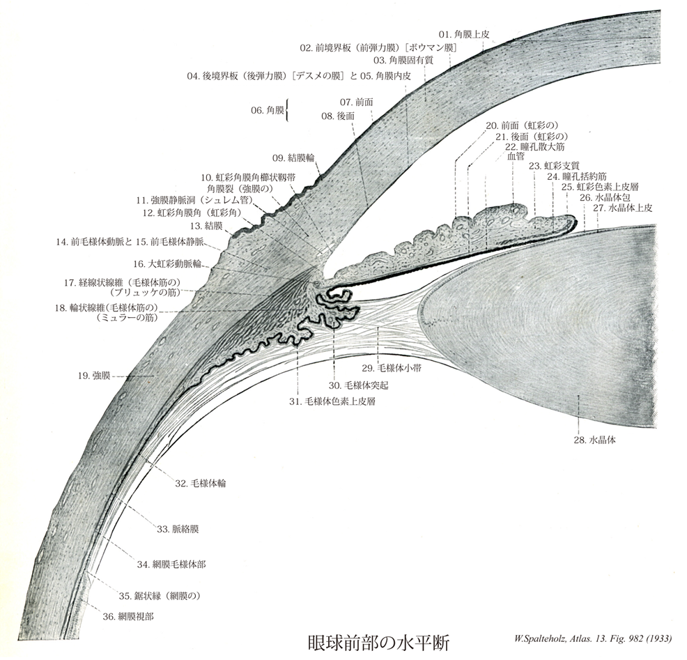
- 982_00【Eyeball眼球 Bulbus oculi】
→(眼球は名前のように球状(直径約25mm・体積約8cm3)で、視覚器の主要部をなす。眼窩脂肪体、眼筋筋膜、眼球鞘などに包まれて眼窩中にあり、前方からは眼瞼により保護されている。また眼筋の働きにより球関節に似た自由度の高い体軸性運動を行う。眼球の内部には前方に眼房水、後方に硝子体が満ちて、12~22mmHgの内圧が保たれる。眼球の形状を規定するため、前極、後極、赤道、経線、外眼球軸(前・後極を結ぶ)、内眼球軸、視軸などを用いる。眼球軸は角膜と水晶体前・後面の曲率中心を通る軸で、網膜面では中心窩と円板の中間を通る。したがって水晶体後面の屈曲率中心と中心窩を結ぶ視軸とは一致しない。眼球壁は組織発生的に、①眼球線維膜(強膜、角膜)、②眼球血管膜(脈絡膜、毛様体筋、虹彩支質、角膜内皮、胎児期の瞳孔膜)、③眼球内膜(網膜視部、毛様体・虹彩色素上皮層)の3層よりなる。①と②は中胚葉、③は神経外胚葉に由来する。内部の水晶体は体表外胚葉、硝子体は中胚葉由来であり、眼瞼・眼球膜、角膜上皮は皮膚の表皮の続きである。)
- 982_01【Corneal epithelium; Anterior corneal epithelium角膜上皮;前角膜上皮 Epithelium anterius corneae】 Approximately five layers of squamous epithelium covering the anterior surface of the cornea. Its surface is very smooth.
→(角膜上皮は角膜縁で眼球結膜につづく重層扁平上皮、結膜と同様に角化しない。無数の知覚神経終末(三叉神経の枝)に含み、刺激に対し鋭敏で、角膜反射をおこす。厚さ~60μm。)
- 982_02Bowman's membrane; Reichert's membrane【Anterior limiting lamina of cornea前境界板;外境界板;前弾力膜;ボウマン膜(角膜の) Lamina limitans anterior; Lamina limitans externa; Lamina elastica anterior】 Basal membrane of the corneal epithelium that is about 10-20 p.thick. It is continuous posteriorly with the substantia propria.
→(ボウマン膜とも呼ばれる。前境界板は角膜上皮と角膜固有質の間の層で、角膜固有質が特殊化したものとされる。表層部は結膜下組織につづく。~10μm。シュワルベ輪ともよばれる。英国の外科医Sir William Bowman (1816-1892)によって記載された。)
- 982_03【Substantia propria of cornea角膜固有質 Substantia propria corneae】 Main, avascular component of the cornea that consists of a lamellar arrangement of connective tissue and a ground substance containing mucopolysaccharides. Corneal transparency is produced by turgescence of the fibers and colloidal fluid distribution.
→(角膜固有質は直線的な膠原線維束からなる厚さ10μmの層が方向を変えて積み重なる。線維間と層間隙は酸性粘液多糖体と組織液に満たされている。固有質は胸膜の続きであり、眼球線維膜の前方部に相当する。)
- 982_04Descemet's membrane【Posterior limiting lamina of cornea後境界板;内境界板;後弾力膜;デスメの膜;(角膜の) Lamina limitans posterior; Lamina limitans interna; Lamina elastica anterior】 Basal membrane of the posterior corneal epithelium. Along its lateral margin it divides into fibers which radiate into the sclera and iris. Aqueous humor passes through the interstices between fibers into the scleral venous sinus.
→(デスメー膜ともよばれる。後境界板は多糖体を含む無構造の~10μmの層。電顕的には固有質につづく細線維層と、角膜内皮の基底膜とに分けられる。1758年。パリの外科医・解剖学者Jean Descemet (1732-1810)が最初に記載したとされるが、イギリスデは同国の外科医Bebedict Duddelが第一発見者とされている。)
- 982_05【Endothelium of anterior chamber of eyeball角膜内皮;後角膜上皮 Epithelium posterius corneae; Endothelium camerae anterioris】 Simple squamous epithelium covering the posterior surface of the cornea.
→(角膜内皮は単層の中皮(中胚葉性上皮)で、虹彩角膜角の内皮を経て、虹彩内皮につづく。眼球血管膜の前方部に相当する。)
- 982_06【Cornea角膜 Cornea】 Transparent, anterior part (1/6) of the eyeball that is anteriorly convex and posteriorly concave. It is 0.9 mm thick in the middle and 1.2 mm thick at the margin.
→(角膜は眼球前方部の透明部分。厚さ約1mm、直径10~12mmの前弯した楕円形の膜で、角膜頂、角膜縁、前および後面を区別する。弯曲度は前面(曲率半径約7.8mm)よりも後面の方が強い。前面は光学的に縦径線が横径線に比してやや強く弯曲し、正視眼ではこの差は水晶体弯曲度の逆の関係により補正されるている。角膜の特徴として、角膜血管が入る辺縁部以外ではまったく血管が存在しない。組織学的に5層が区別される。①角膜上皮、②前境界板(Bowman膜)、③角膜固有質、④後境界板(Descement膜)、⑤角膜内皮)
- 982_07【Anterior surface of cornea前面(角膜の) Facies anterior corneae】 Surface of the cornea facing the outside air.
→(角膜の前面は空気に接する角膜前面。)
- 982_08【Posterior surface of cornea後面(角膜の) Facies posterior corneae】 Surface of the cornea facing the anterior chamber.
→(角膜の後面は前眼房に接する角膜後面。)
- 982_09【Conjunctival ring結膜輪 Anulus conjunctivae】 Transition of the conjunctival epithelium into the epithelium of the anterior surface of cornea.
→(結膜輪は結膜と角膜辺縁の接合部の幅の狭い輪で結膜上皮から角膜上皮への移行部。)
- 982_10【Pectinate ligament of iridocorneal angle虹彩角膜角櫛状靱帯;小柱網;櫛状靱帯(強膜の) Ligamentum pectinatum anguli iridocornealis】
→(虹彩角膜角櫛状靱帯と虹彩角膜角隙をより的確に表すために“小柱網”が用いられる。虹彩支質に移行する後方部をブドウ膜部、強膜棘の内側から角膜内皮に移行する前方部を強膜部という。)
- 982_11Schlemm, Canal of【Scleral venous sinus; Schlemm's canal; Canal of Schlemm強膜静脈洞;シュレム管 Sinus venosus sclerae】
→(シュレム管とも呼ばれる。強膜静脈洞は輪状に走る静脈性血管。内側は虹彩角膜角櫛状靱帯により境される。中断もしくは重複することあり。前眼房から眼房水を受け入れる。Schlemm, Friedrich (1795-1858) ドイツの解剖学者1830年に角膜と強膜の間にある環状小静洞について記載(""Arteriarum capitis superficialium icon nova"", Berolini, J.W. Boike)。)
- 982_12【Iridocorneal angle虹彩角膜角;虹彩角 Angulus iridocornealis; Angulus iridis】 Angle between the iris and cornea. It contains the trabecular tissue, which allows drainage of aqueous humor through its interstices into the scleral venous sinus.
→(虹彩角膜角は虹彩角膜角櫛状靱帯を有し、この靱帯の隙間を通って眼房水は強膜静脈洞へ入る。)
- 982_13【Conjunctiva結膜 Tunica conjunctiva】 Membrane covering the inner surface of the eyelids where it is composed of stratified (dual-layered or multilayered) columnar epithelium with goblet cells and a loosely organized, well-vascularized lamina propria containing numerous cells. It reflects at the conjunctival fornix onto the eyeball, covering it with stratified squamous epithelium as far as the corneal margin.
→(結膜は上・下眼瞼の内面(眼瞼結膜)と眼球前面(眼球結膜)をおおう粘膜である。眼球結膜は厚く不透明で血管に富み、表面に多数の乳頭をもつ。後眼瞼縁で結膜は眼瞼線分泌間の上皮にめがしらの半月ヒダの結膜は涙湖の底をつくり、涙点、涙管を経て鼻涙管粘膜上皮につづく。上眼瞼外側核の円蓋には6~12本の涙腺管が開く。上・下の結膜円蓋を経て、眼瞼結膜が眼球結膜に移行する。眼球結膜はゆるやかに強膜表面をおおい、薄く透明で乳頭を欠き血管分布に乏しく結膜輪で角膜上皮に移行する。円蓋の結膜には小形の杆状胞状腺(結膜腺、クラウゼ線)がある。)
- 982_14【Anterior ciliary arteries前毛様体動脈 Arteriae ciliares anteriores】 Arteries arising from the anterior muscular arteries and passing through the sclera to the choroid and ciliary body before opening at the major circulus arteriosus of iris.
→(前毛様体動脈は角膜縁のやや後方で眼球壁に入り、強膜上動脈、前結膜動脈および後結膜動脈を分岐する。角膜縁近くで強膜を貫いて進入し、大虹彩動脈輪と連絡する。)
- 982_15【Anterior ciliary veins前毛様体静脈 Venae ciliares anteriores】 Veins accompanying the anterior ciliary artery that convey blood from the ciliary body to the veins of the ocular muscles at their beginning.
→(前毛様体動脈は角膜縁のやや後方で眼球壁に入り、強膜上動脈、前結膜動脈および後結膜動脈を分岐する。)
- 982_16【Major circulus arteriosus of iris; Major arterial circle of iris大虹彩動脈輪 Circulus arteriosus iridis major】 Vascular circle with radiating branches formed by anastomoses between the long and short posterior ciliary arteries.
→(大虹彩動脈輪は長および短後毛様体動脈管の吻合による輪状の血管系。放射状に枝がでる。)
- 982_17Bücke's muscle【Meridional fibres of ciliary muscle経線状線維;経線線維(毛様体筋の);ブリュッケの筋 Fibrae meridionales (Musculus ciliaris)】 Larger group of fibers running meridionally that mostly arise from the scleral spur and pass along the choroid to the sclera.
→(ブリュッケ筋とも呼ばれる。毛様体筋のうちの経線状に走る平滑筋線維。筋の大部分を占める。前方、後境界板のところは虹彩角膜角櫛状靱帯へ、後方は脈絡膜へつく。眼球の毛様体内部に含まれる。オーストラリアの生理学者Ernest W. von Bruecke (1819-1892)の名を冠する。この筋はアイルランドの外科医Sir Philip Crampton (1777-1892)の名を冠する。この筋はアイルランドの外科医Sir Philip Crampton (1777-1858)のなをとってCrampton's muscleとも呼ばれる。なお、毛様体の輪状平滑筋はミュラー筋(Mueller's muscle)と呼ばれている。)
- 982_18Müller's muscles【Circular fibres of ciliary muscle輪状線維(毛様体筋の) Fibrae circulares (Musculus ciliaris)】 Anular inner muscle layer.
→(ミュラー筋ともいう。眼球の毛様体内部にある輪状に走る平滑筋線維。ドイツの解剖学者Heinrich Franz Mueller (1820-1864)によって報告された。経線方向に走る平滑筋線維はブリュッケ筋Brucke's muscleという。)
- 982_19【Sclera強膜 Sclera】 Membrane of the eyeball composed of interwoven collagen fibers. It has a bluishwhite appearance and is visible through the conjunctiva.
→(眼球の形状を保つ強靱な膠原線維組織層。角膜となっている前部6分の1を除いた部分。前方では隔膜固有質に、後方では篩板から視神経外鞘を経て脳硬膜に、それぞれつづいている。強膜と角膜を合わせて眼球線維膜という。強膜の厚さは眼球後極で~1.0mm、前部で~0.6mm、赤道で~0.4mmである。視神経線維束を通す篩板は後極の内側3.5mm、視神経乳頭の直後方にあたる。視神経は~数十本の掌側としてこれを通る。渦静脈、長・短毛様体動脈および神経が強膜を貫く。強膜はは外から内へ、①強膜上皮、②強膜固有質、③強膜褐色板の3沿うよりなる。虹彩角膜角に沿って強膜固有質が内方へ皮厚し(強膜距)毛様体筋腱により貫かれる。この部の直前に輪状に走る強膜静脈洞(Schlemmn管)があり、眼房水は虹彩角膜間隙(Fontana腔)からこれを通って渦静脈に排出される。角膜縁をとり膜浅い強膜溝の深層にこれらの構造がある。眼球前部の強膜上板毛細血管網に富み、その炎症性変化を臨床的に「網膜充血」という。強膜前部は眼球結膜、後部は眼球鞘(Tenon鞘)によりおおわれる。内面は脈絡外隙を間に脈絡外板に接する。)
- 982_20【Anterior surface of iris前面(虹彩の) Facies anterior iridis】 Anterior surface of the iris facing the anterior chamber.
→(前眼房へ対する面。(Feneis))
- 982_21【Posterior surface of iris後面(虹彩の) Facies posterior iridis】 Posterior surface of the iris facing the posterior chamber.
→(虹彩の後面は後眼房へ対する面。)
- 982_22【Dilator pupillae muscle瞳孔散大筋 Musculus dilator pupillae】 Thin layer of smooth muscle whose cells are mainly oriented radially. It is innervated by sympathetic nerve fibers via the carotid plexus.
→(放射状に走るうすい平滑筋線維層。神経支配は頚動脈神経叢からの交感神経。(Feneis))
- 982_23【Stroma of iris虹彩支質 Stroma iridis】 Vascular framework of the iris interspersed with pigmented connective-tissue cells. Its anterior and posterior portions are denser, with fine fibers in between.
→(虹彩支質は色素を持つ結合組織細胞の混じった、血管に富む基質。前および後部は線維が多く、中間部は少ない。(Feneis))
- 982_24【Sphincter pupillae; Sphincter muscle of pupil瞳孔括約筋 Musculus sphincter pupillae】 Meshwork of spiraling muscle fibers. In the dilated pupil its long axes run nearly parallel to the pupillary margin. It is innervated by parasympathetic fibers from the oculomotor nerve.
→(瞳孔括約筋はラセン状に走る筋線維よりなる網工。その長軸は、散大した瞳孔では瞳孔縁とほぼ平衡である。神経支配は動眼神経中の副交感性線維。瞳孔に近い色素上皮層前面に束をつくるのみならず、一部は瞳孔縁から後面にまで及んでいる。)
- 982_25【Pigmented layer of iris虹彩色素上皮層;虹彩色素層 Stratum pigmenti iridis】
→(網膜虹彩部は虹彩の後面を縁どる上皮層で、網膜毛様体部と同様に、自由表面を互いに向かい合わせた2層の上衣細胞からできている。虹彩支質に接する前面の上皮は眼杯外板に由来する虹彩色素上皮層であり、後眼房に向かう後面の上皮は眼杯内板に由来するもので、狭義の網膜虹彩部と呼ばれ、これは瞳孔縁のところで反転して虹彩色素上皮に移行する。虹彩色素上皮層の細胞は特異な細胞で、核のあるレベルを境としてそれより後方、すなわち自由表面側の細胞質は大量のメラニン顆粒を含み、単層立方上皮様配列を示すが、核よりも前方、すなわち基底側細胞質はメラニン顆粒を含まず、細長い紡錘形の突起となり、虹彩支質と色素上皮層の接着面に平行に、虹彩のつけねから瞳孔縁に向かって走る。これが瞳孔散大筋そのものである。)
- 982_26【Capsule of lens水晶体包;水晶体被膜 Capsula lentis】 Crystal-clear membrane up to 15 (im thick covering the lens and its epithelium. It is thicker at the anterior pole than at the posterior pole. It gives attachment to zonular fibers.
→(水晶体包は透明な15μmまでの厚さの被膜。上皮も含めて水晶体全面を被う。前極では後極よりも厚い。)
- 982_27【Lens epithelium水晶体上皮 Epithelium lentis】 Epithelium of the lens that extends to the equator of the lens. It is derived during embryonic development from the anterior wall epithelium of the lens vesicle.
→(水晶体上皮は水晶体前面にあり、水晶体赤道にまで達する上皮。発生学的には水晶体小胞の前壁上皮に由来する。)
- 982_28【Lens水晶体 Lens; Lens crystallina】 Lens suspended by the ciliary zonule between the pupil and vitreous body. It measures 9-10 mm in diameter and is about 4 mm thick.
→(水晶体は虹彩の後方、硝子体の前方に位置し、双凸面レンズ構造をもつ。赤道直径約9mm、水晶体軸(前、後極を結ぶ直線)3.7~4.4mm、前面弯曲度から8mm、後面弯曲度~6mm、屈折率1.36(中央部)~1.42(辺縁部)。水晶体は無色透明な水晶体包(前面で厚く、後面で薄い粘液多糖体層で、水晶体上皮の基底膜が発達したもの)におおわれる水晶体質よりなる。水晶体質はより軟らかい上皮と硬い核に分かれやすく、胎児では雌で水晶体包に切れ目をいれるとはじけるように裂ける。生体では前、後極から発する数本の水晶体放線がわずかに認められ、胎児では前後両面放線がわずかに認められ、胎児では前後両面に、たがいに120°に交わる3本の放線(前面逆Y字、後面正Y字形)を示す。水晶体の構成要素は水晶体線維で、発生初期の単層の水晶体胞の後壁の細胞のみが著しく長大化したものである。前面に層単層の水晶体上皮は水晶体胞前壁の原型を保つ。赤道より後面にいくにしたがい長細い六角形の水晶体線維の束へと移行する。胎児期の放線は水晶体線維束の付着点をなす中隔に一致し、前極からおこる線維は後面の赤道近くの最寄りの放線に、前面赤道近くの中隔よりおこる線維は後極へ向かう。水晶体線維は緊密かつ生前と配列するが、微絨毛を出して細い細胞管腔を確保し、水および代謝物質の移送路を形成する。生体の水晶体には血管の神経の分布が認められない。胎児の水晶体包は硝子体動脈により養われるが、妊娠末期に道動脈が閉鎖する。老年者では前後面の弯曲度が減って扁平となり、黄白色を帯びる傾向にある。この変化が進行したものを白内障cataractという。全体の25%を占める水晶体蛋白はα-およびβ-クリスタリンと不溶性アルブモイドよりなり、そのほかにグルタチン、ビタミンCなどが含まれる。)
- 982_29Zinn, Zonule of【Ciliary zonule毛様体小帯;水晶体小帯 Zonula ciliaris; Apparatus suspensorius lentis】 Suspensory apparatus of zonular fibers and spaces encircling the equator. It is composed of system of radiating fibers of varying lengths and the folds between them.
→(チン小帯ともよばれる。水晶体被膜を毛様体突起に固定する索で、個々の線維を小帯線維という。毛様体突起の比較的後部よりおこる線維は、水晶体赤道の前方に、前方よりおこるものは、後方に付着する。したがって一部線維が交叉する。小帯線維の間に残る小帯隙を経て、後眼房から前眼房へ眼房水が移動する。ドイツの解剖学者Johann Gottifried Zinn (1727-1759)による。)
- 982_30【Ciliary processes毛様体突起;大突起(毛様体の) Processus ciliares; Processus majores】 Between 70 and 80 radiating folds containing numerous capillaries measuring 0.1-0.2 mm wide, 1 mm high, and 2-3 mm long. Their epithelium produces aqueous humor.
→(毛様体突起は放射状に配列し、毛細血管に富むヒダ。70~80本あり、幅0.1~0.2mm、高さ1mm、厚さ2~3mmである。この上皮が眼房水を産出する。)
- 982_31【Pigmented layer of ciliary body毛様体色素上皮層;毛様体色素層 Stratum pigmenti corporis ciliaris】
→(網膜毛様体部は自由表面を向かい合わせた2層の上皮細胞から出来ている。外層は眼球外板に由来し、網膜色素上皮層に直接続くもので、毛様体色素上皮層と呼ばれ、大量のメラニン顆粒を含む単層円柱上皮である。これと外側を囲む血管層の間にはブルッフ膜につづく基底膜が存在する。内層は眼杯内板に由来するもので、毛様体上皮とも呼ばれる単層円柱ないし立方上皮である。細胞はメラニン顆粒を含まず、細胞体はHE染色では明るい桃色に染まる。後眼房に向かうこの細胞の表面は、発生過程から明らかなように、自由表面ではなくて基底面であり、基底膜に相当する内境界膜で裏打ちされている。おの内境界膜に小帯線維が付着する。毛様体上皮は眼房水を分泌するものと考えられているが、その分泌の機序はまだよくわかっていない。)
- 982_32【Orbiculus ciliaris毛様体輪 Orbiculus ciliaris】 Circular zone between the corona ciliaris and ora serrata that is covered with ciliary plicae.
→(毛様体輪は毛様体冠と鋸状縁の間にある輪状帯。毛様体ヒダがある。(Feneis))
- 982_33【Choroid脈絡膜 Choroidea; Chorioidea】 Portion situated between the retina and sclera.
→(脈絡膜は網膜視部の外側に接してこれを包んでおり、内側から外側に向かって、①基底膜、②脈絡毛細血管板、③血管板、④脈絡上板の4層に分けられる。)
- 982_34【Ciliary part of retina網膜毛様体部 Pars ciliaris retinae】 Part situated on the posterior surface ciliary body. It is composed of simple, cuboidal, pigmented epithelium.
→(網膜毛様体部は自由表面を互いに向かい合わせた2層の上皮細胞からできている。外層は眼球外板に由来し、網膜色素上皮層に直接つづくもので、毛様体色素上衣層と呼ばれ、大量のメラニン顆粒を含む単層円柱上皮である。これと外側を囲む血管層の間にはブルッフ膜につづく基底膜が存在する。内層は眼杯内板に由来するもので、毛様体上皮とも呼ばれる単層円柱内し立方上皮である。細胞はメラニン顆粒を含む、胞体はHE染色では明るい桃色に染まる。後眼房に向かうこの細胞の表面は、発生過程からあきらかなように、自由表面ではなくて基底面であり、基底膜に相当する内境界膜で裏打ちされている。この内境界膜に小帯線維が付着する。毛様体上皮は眼房水を分泌するものと考えられている。)
- 982_35【Ora serrata retinae鋸状縁(網膜の) Ora serrata retinae】 Serrated border between the light-sensitive and light-insensitive portions of the retina.
→(網膜の鋸状縁は網膜視部と毛様体部との間の鋸歯状の境界。)
- 982_36【Optic part of retina網膜視部;視部(網膜の) Pars optica retinae】 Portion of the retina capable of transforming light impulses into nerve impulses. It borders posteriorly with the ora serrata.
→(網膜視部はHE染色標本でみると、内側から外側へ向かって規則正しい10層の層構造を示す。)