Spalteholz HANDATLAS DER ANATOMIE DES MENSCHEN VON WERNER SPALTEHOLZ
メニューは解剖学(TA)にリンクしてあります。図の番号をクリックすると下記の説明へ、右側の用語をクリックすると解剖学(TA)にジャンプします。
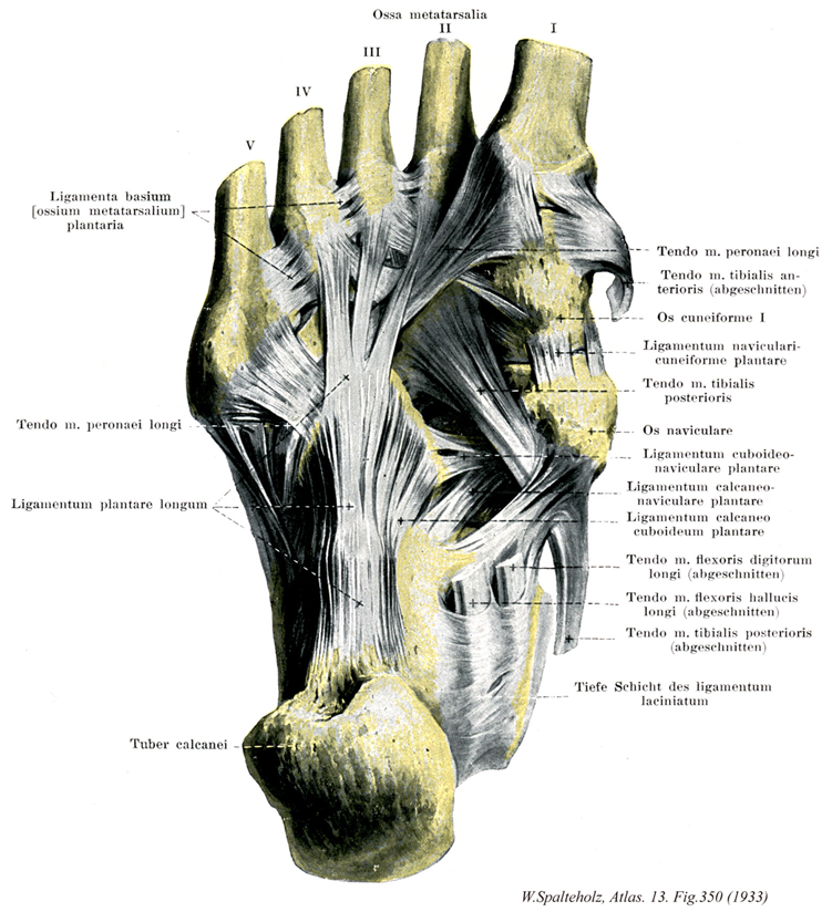
350


- 350_00【Joints of foot足の関節;足関節 Articulationes pedis】
→(距腿関節をも含めて、足根骨、中足骨および足の指骨の間に生ずるすべての関節を総称していう。狭義では距腿関節のみを指す。(解剖学辞典:河西達夫))
- 350_01【Metatarsals; Metatarsal bones [I-V]中足骨[1-5] Ossa metatarsi; Ossa metatarsalia [I-V]】 The five metatarsal bones.
→(中足の骨格で5個の長骨からなる。内側から順に第1・第2・第3・第4および第5中足骨という。第1・第2および第3中足骨はそれぞれ内側・中間および外側楔状骨の遠位に、第4および第5中足骨は立方骨の遠位にある。長さは中手骨より長い。第1中足骨が最も短く、第2中足骨が最も長い。第3・第4および第5中足骨の順に短くなる。おのおのの中足骨を近位端の底、中央部の体、遠位端の頭に分ける。底は太く厚く、第1・第2および第3中足骨にはそれぞれ内側・中間および外側楔状骨に対する、第4と第5中足骨には立方骨に対する関節面がある。第1中足骨を除く中足骨と足根骨の関節を連ねる線は、内側前方から外側後方へ走る。第2・第3・第4および第5中足骨底では相対する側面に関節面があるが、第1中足骨底にはない。また第2中足骨底の側面には内側および外側楔状骨に対する関節面がある。第1中足骨底の足底面に第1中足骨粗面がある。第5中足骨底の外側は強く張り出して第5中足骨粗面をなし、皮下で触知できる。体は不正三角柱で、第1中足骨では太いが、他は左右から圧迫された形をし、頭へいくほど細くなる。また長軸方向で背側に凸弯している。頭は側面を切り取った球状で、細い頚部がある。頭の足底面には内および外側に、それぞれちいさな隆起がある。)
- 350_01a【First metatarsal bone; 1st metatarsal bone第1中足骨 Os metatarsale I】
→()
- 350_01b【Second metatarsal bone; 2nd metatarsal bone第2中足骨 Os metatarsale II】
→()
- 350_01c【Third metatarsal bone; 3rd metatarsal bone第3中足骨 Os metatarsale III】
→()
- 350_01d【Fourth metatarsal bone; 4th metatarsal bone第4中足骨 Os metatarsale IV】
→()
- 350_01e【Fifth metatarsal bone; 5th metatarsal bone第5中足骨 Os metatarsale V】
→()
- 350_02【Plantar metatarsal ligaments底側中足靱帯;底側中足骨底靱帯 Ligamenta metatarsalia plantaria; Ligamenta basium ossium metatarsi plantaria】 Fibrous bands connecting the plantar surfaces of the bases of the metatarsals.
→(底側中足靱帯は各中足骨底の底面を結ぶ。)
- 350_03【Fibularis (peroneus) longus tendon長腓骨筋腱 Tendo musculus peroneus longus; Tendo musculus fibularis longus】
→()
- 350_04【Long plantar ligament長足底靱帯 Ligamentum plantare longum】 Firm band that passes from the calcaneus just anterior to the calcaneal tuberosity to the cuboid and the bases of metatarsals II-V. It supports the longitudinal arch of the foot.
→(長足底靱帯は足底の靱帯のうち最も表層にありまた最も長い。そのほかの底側足根靱帯はこれにより下方から被われ、その間に粗な結合組織が介在する。距骨隆起の下面から起こって前方に広がり、その深層の線維は立方骨の長腓骨筋腱溝の後の立方骨粗面に着く。浅層の線維は同腱の表面を越えて少なくも3束に分かれて中足骨底に着く。)
- 350_05【Calcaneal tuberosity踵骨隆起 Tuber calcanei】 Tuberosity on the posterior aspect of the calcaneus.
→(踵骨の後半部は大きな骨塊となって後方に飛び出している。この部分は踵骨隆起と呼ばれ、いわゆるかかとの主要部を成している。その後面には表面にギザギザした稜線が横に走っているが、ここはアキレス腱がつく場所である。)
- 350_06【Tibialis anterior tendon前脛骨筋腱 Tendo musculus tibialis anterior】
→()
- 350_07【Medial cuneiform bone内側楔状骨;第一楔状骨 Os cuneiforme mediale; Os cuneiforme primum】 Wedge-shaped bone between the navicular and the first metatarsal. The base of the wedge is directed inferiorly.
→(舟状骨と第一中足骨の間に位置する。その楔形の底面は下方を向いている。3つの楔状骨の中で内側楔状骨は飛び抜けて大きい。)
- 350_08【Plantar cuneonavicular ligaments底側楔舟靱帯 Ligamenta cuneonavicularia plantaria; Ligamentum navicularicuneiforme plantare】 Group of ligaments that connect the navicular with the cuneiform bones in front of it.
→(足底楔舟靱帯は舟状骨と内側・中間・外側楔状骨の底足面を結ぶ靱帯で、下面は後脛骨筋の腱から放散した線維によって補強される。)
- 350_09【Tibialis posterior tendon; Tendon of tibialis posterior muscle後脛骨筋腱 Tendo musculus tibialis posterior】
→()
- 350_10【Navicular bone舟状骨[足の] Os naviculare】 Bone situated medially between the head of the talus and the three cuneiform bones.
→(足の舟状骨は中心足根骨に属し、足根の内側で距骨と楔状骨の間にある。前後に扁平な骨で、背側で凸面、足底側で凹面となっている。前面に3個の凸面の関節面があり、それぞれ内側楔状骨・中間楔状骨・外側楔状骨と関節する。後方は距骨頭に対する関節窩をなす。内側面は粗面状で下方に突出し舟状骨粗面をなす。皮下で触知できる。外側面で立方骨関節することも多い。底側面で舟状骨粗面に近い部に後脛骨筋腱の深層のものが通る浅い斜めの溝がある。ラテン語のNavisの縮小形Navicula(小舟)に由来する。)
- 350_11【Plantar cuboideonavicular ligament底側立方舟靱帯 Ligamentum cuboideonaviculare plantare】
→(底側立方舟靱帯は立方骨と舟状骨の底足面を結ぶ靱帯で横走する。)
- 350_12【Plantar calcaneonavicular ligament; Spring ligament底側踵舟靱帯;スプリング靱帯 Ligamentum calcaneonaviculare plantare】 Ligament that passes from the sustentaculum tali to the plantar and medial surfaces of the navicular, augmenting the articular surface for the head of the talus.
→(底側踵舟靱帯は踵骨の載距突起の前縁下面と内側縁から起こり、舟状骨の底面、内側縁と上面の内側部を結ぶ幅の広い厚い靱帯で、距踵舟関節包の一部となる。一部は同関節腔内に露出して距骨頭の下面に接する凹面となり、線維軟骨化している。この靱帯の内側部は内側靱帯脛舟部の線維と合し、それによって補強されている。上面は距踵舟関節の関節腔内に露出して、距骨頭を下方から支える関節面を形成し、この部分は線維軟骨化して滑液膜におおわれている。この靱帯の下方は後脛骨筋の腱によって支えられ、その内側縁は距腿関節の内側靱帯の前部と癒合している。足弓を支持する役割をもち、距骨と舟状骨の間に張って足弓の頂点の位置にある。この靱帯の無力化は、距骨頭が体重の負荷によって前内下方に押されて扁平足の原因となる。またこの靱帯は弾性線維を含んで足弓に弾性を与え、spring ligamentともよばれる。)
- 350_13【Plantar calcaneocuboid ligament; Short plantar ligament底側踵立方靱帯;短足底靱帯 Ligamentum calcaneocuboideum plantare】 Shorter fibers of the long plantar ligament.
→(底側踵立方靱帯は踵骨下面の前方部(踵骨結節)から起こり、前方に放散して立方骨粗面に着く。)
- 350_14【Flexor digitorum longus tendon長趾屈筋腱 Tendo musculus flexor digitorum longus】
→()
- 350_15【Flexor hallucis longus tendon長母趾屈筋腱 Tendo flexor hallucis longus; Tendo musculus flexor hallucis longus】
→()
- 350_16【Flexor retinaculum of foot屈筋支帯[足の];破裂靱帯 Retinaculum musculorum flexorum pedis; Ligamentum laciniatum】 Multilayered band situated over the long flexor tendons passing from the medial malleolus to the calcaneus. Its superficial portion invests the tibial nerve and posterior tibial artery and veins. Its deep portion forms an osteofascial canal with compartments containing the posterior tibial flexor muscles, flexor digitorum longus, and flexor hallucis longus.
→(足の屈筋支帯は下腿筋膜の厚くなったもので、内果の下部から扇状に広がって、前部は舟状骨に後部は踵骨につき中間部は足底腱膜に移行する。屈筋支帯は後脛骨筋と長趾屈筋の腱を被い、またその間の隔壁を骨に送ったのち、載距突起についてさらに長母指屈筋腱溝を被う深葉と、脛骨神経および後脛骨動静脈を被う浅葉とに分かれる。)