Spalteholz HANDATLAS DER ANATOMIE DES MENSCHEN VON WERNER SPALTEHOLZ
メニューは解剖学(TA)にリンクしてあります。図の番号をクリックすると下記の説明へ、右側の用語をクリックすると解剖学(TA)にジャンプします。
497 


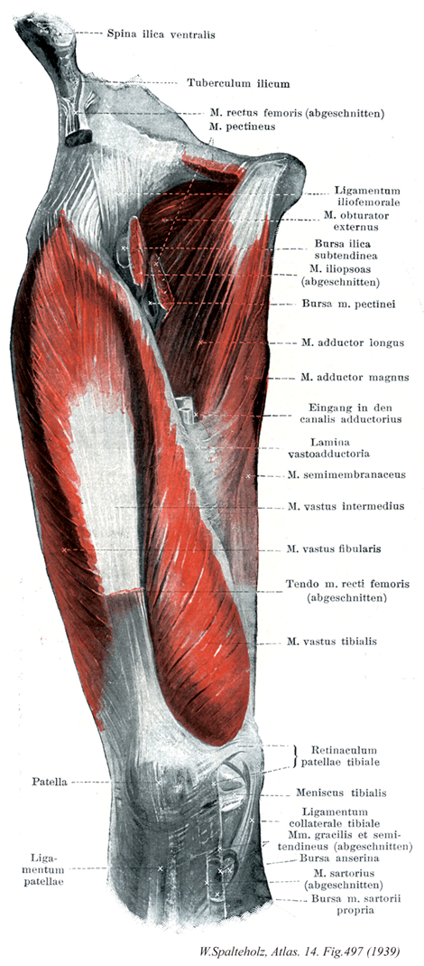
- 497_01【Patella膝蓋骨 Patella】 The kneecap, which is embedded in the tendon of the quadriceps femoris muscle.
→(膝関節の前面にあり、尖端を下方に向けた扁平な栗の実によく似た骨で、幅広い上端部が膝蓋骨底で、尖った下端部が膝蓋骨尖である。大腿四頭筋腱中に発生した種子骨とみなされ、上縁には大腿直筋と中間広筋の内側縁には内側広筋の、外側縁には外側広筋のそれぞれの腱が付着する。前面は凸面状で、大腿四頭筋腱による縦に走る小隆起を伴う粗面をなし、小血管孔がある。後面には、上方の広い卵形の平滑な面と、下方の小さい逆三角形の粗な面がある。平滑な面は大腿骨の膝蓋面に対する関節面をなし、中央部にある縦方向の隆起によって小さい内側部と大きい外側部に分けられる。下方の粗面の下端には膝蓋靱帯が付着するが、粗面の上方部には脂肪組織が入り、脛骨と膝蓋骨とを隔てる。ラテン語のPatera(皿・円板状の)の縮小形。)
- 497_02【Patellar ligament膝蓋靱帯;膝蓋腱 Ligamentum patellae】 Ligamentous continuation of the tendon of the quadriceps femoris muscle that passes from the apex of the patella to the tibial tuberosity. It is 2-3 cm wide and about 0.5 cm thick.
→(膝蓋腱と膝蓋靱帯は同義語。本体は大腿四頭筋の停止腱であるから、膝蓋腱という方が理論的には正しいが、膝蓋骨の下部から起こって脛骨粗面に着く強靱な線維束を膝蓋靱帯といい、膝蓋骨に着く上端が幅広く、下部が細い。また膝関節包を補強する意味から膝蓋靱帯という名が採用されている。また膝蓋腱とう語も慣用されている。)
- 497_03【Anterior superior iliac spine; Iliospinale anterius上前腸骨棘;前腸骨棘 Spina iliaca anterior superior; Spina ilica ventralis】 Bony projection at the anterior border of the iliac crest giving origin to the sartorius muscle.
→(腸骨稜の前端は鈍円な突起として大きく突出し、上前腸骨棘として体表上からもよく触れる。大腿筋膜張筋および縫工筋が起こる。)
- 497_04【Tuberculum of iliac crest腸骨結節(腸骨稜の) Tuberculum iliacum】 Palpable projection on the outer lip about 5 cm posterior to the anterior superior iliac spine at the junction of the anterior gluteal line and the iliac crest.
→(上前腸骨棘の後方5cmにあり、前臀筋線と腸骨稜とが出会う部位にある外唇の隆起。触れることができる。)
- 497_05【Rectus femoris muscle大腿直筋 Musculus rectus femoris】 Two-headed muscle. Also flexes the hip joint. Contributes fibers to the lateral and medial patellar retinacula.
→(大腿直筋は大腿前側の筋で大腿四頭筋の中央浅層にある。羽状筋の大腿直筋は下前腸骨棘から起こる直頭と寛骨臼上縁や股関節から起こる反転頭からなる。起始腱中心部からの線維は膝蓋骨の上縁に終わるが、膝蓋骨前面を通って膝蓋靱帯となる。両側方の線維は膝蓋骨の両側へ放散し、膝蓋支帯になる。)
- 497_06【Pectineus muscle恥骨筋 Musculus pectineus】 o: Pectineal line of pubis. i: Pectineal line of femur, linea aspera. Flexion, adduction, and medial rotation at the hip. I: Femoral and obturator nerves.
→(恥骨筋は腸恥隆起-恥骨結節間の恥骨上肢から起こり、大腿骨の恥骨筋線に停止する。この筋はもとももと腸腰筋群と同じ原基に由来する。本筋の構成に内転筋群がどの程度関わるかには個体差がある。恥骨筋は腸骨筋膜の延長部分である恥骨筋膜におおわれ、腸腰筋とともに腸恥窩には大腿動静脈が通る。)
- 497_07Bertin's ligament; Bigelow's ligament【Iliofemoral ligament腸骨大腿靱帯 Ligamentum iliofemorale】 Strong anterior band that extends from the anterior inferior iliac spine to the intertrochanteric line. It travels in two main directions.
→(腸骨大腿靱帯は下前腸骨棘および寛骨臼上縁から起こり、拡がって転子間線に至る三角形の強い靱帯で、関節包の前面と上面を補強する。しかし転子間線に近い中央部はやや弱いので、強い線維束となる部は逆Y字形となる。特にその外上部は、上方では下前腸骨棘に着く大腿直筋腱の線維、下方では大転子に着く小臀筋腱の線維が表面から加わり、股関節包靱帯のなかでも最大の部分となる。腸骨大腿靱帯は靱帯最大の靱帯で、上半身が股関節より後へ傾くのを防ぐバンドの役目をしている。つまり直立のバランスをとるためには、腸腰筋と共に不可欠のものである。なお上半身が股関節で前へ傾くのを防ぐものとしては、大腿筋が最も重要である。腸骨大腿靱帯の三角筋は両側辺が厚くて中央部が薄いので、英仏ではV字靱帯、またはY字靱帯Y-shaped ligamentと呼んでいる。)
- 497_08【Obturator externus muscle; External obturator muscle外閉鎖筋 Musculus obturator externus】 o: External surface of obturator membrane and surrounding area, i: Trochanteric fossa. Lateral rotation and adduction at the hip joint. I: Obturator nerve.
→(外閉鎖筋は閉鎖膜外面および閉鎖孔内下方の骨縁に起こる。腹内側へ走り、股関節の背側で大腿骨頚と頭をまわりこんでから錐状の停止腱に移行し、腹外側へ向かって転子窩に停止する。)
- 497_09【Subtendinous bursa of iliacus腸骨筋腱下包;腸骨腱下包 Bursa subtendinea iliaca; Bursa ilica subtendinea】 Bursa situated between the lesser trochanter and the tendon of insertion of the iliopsoas.
→(腸骨筋の腱下包は小転子と腸腰筋停止腱との間にある。)
- 497_10Hyrtl's muscle【Iliopsoas muscle腸腰筋 Musculus iliopsoas】 Muscle comprising the psoas major and iliacus. i: Lesser trochanter. Most important anterior flexor of the leg. Trunk flexion, lateral rotation.
→(腸骨筋と大腰筋からなる複合筋で、共同腱によって大腿骨小転子の前面に停止する。内側部(大腰筋、長線維、大きな挙上作用を持つ)は第12胸椎および第1から4腰椎外側面から起こる深層と大腰椎の肋骨突起から起こる浅層から成る(この両層間に腰神経層の大部分が入る)。外側部(腸骨筋、多数の線維を持ち、大きな力として作用する)は腸骨窩を埋める。骨盤外の起始は股関節の関節包から起こる(関節包張筋)。大腰筋も腸腰筋もともに筋裂孔を(大腿神経とともに)通って骨盤から出る。大腿骨頚の内側をまわりこみ、関節包とは腸恥包で隔てられる。腸恥包は時々関節腔につながる。しばしば他の滑液包、腸骨筋腱下包が小転子と腸腰筋腱の間にある。頭側では薄い大腰筋の筋膜は尾側では厚くなり、腸骨筋の筋膜と合流して鼡径靭帯外側部に強く結びつくとともに、強い結合組織膜として腸腰筋の停止までをおおう。第3の寛骨内筋、小腰筋は人で常在しない。その起始は第12胸椎と第1腰椎である。長い腱は大腰筋上を尾側へ向かい、腸腰筋膜や、特に腸恥骨弓に放散する。)
- 497_11【Pectineus musculus bursa恥骨筋包;恥骨筋嚢 Bursa musculus pectineus】
→()
- 497_12【Adductor longus muscle長内転筋 Musculus adductor longus】 o: Near the pubic symphysis. i: Medial lip of linea aspera. Adduction, lateral rotation, and flexion at the hip joint. I: Obturator nerve.
→(長内転筋は恥骨結合の線維軟骨および恥骨稜直下の狭い領域から長い腱として起こる。遠位で広くなり、薄い幅広の腱となって粗線(内側唇)の中1/3に停止する。停止腱の遠位縁をつくる筋束は広一内転筋板の形成に多少なりとも参加し、内転筋管の入り口をかたちづくる。)
- 497_13【Adductor magnus muscle大内転筋 Musculus adductor magnus】 o: Inferior pubic ramus, ramus of ischium. i: Medial lip of linea aspera and, via a long tendon, the medial epicondyle. Adduction, lateral rotation, and extension at the hip joint. I: Obturator and tibial nerves.
→(大内転筋は内転筋群の中で最強、人体中最大の筋の1つである。他の内転筋の背側、恥骨下枝および隣接する坐骨枝から坐骨結節までを起始とする。他の起始をもつ部分はほとんどが粗線内側唇に筋性停止する。)
- 497_14Hunter's canal【Adductor canal内転筋管 Canalis adductorius】 Passageway that is formed by the adductor magnus, vastus medialis, and anteromedial intermuscular septum. It ends at the adductor hiatus.
→(ハンター管および縫工筋下管ともよばれる。大腿のほぼ中央の高さにある筋膜性の管で、上方は大腿三角の先端よりつづき、下方は内転筋腱裂孔に開く。外側壁は内側広筋、内側壁は大内転筋と長内転筋によってつくられ、また前壁は、縫工筋の深層をおおう厚い筋膜(広筋内転筋板(INA))が、内側広筋と大内転筋および長内転筋の間に張ることによってつくられる。このなかを大腿動静脈と伏在神経が走る。内転筋管の入口より上の方でも、既に大腿動静脈は長内転筋・縫工筋・内側広筋で囲まれたトンネルを通っている。そしてこのトンネル(筋性内転筋管muscular adductor canalともいう)を上方に場所が、大腿三角の底を作っている腸恥窩である。Williamの弟、John Hunter (1728-1793)によって記載された。精巣下降に関わる精巣導帯にも名を残す。かれの業績はロンドンのRoyal College of SurgeonsのHunter Museumにおいて見ることができる。)
- 497_15【Vastoadductor membrane; Adductor fascia広筋内転筋板;広筋内転筋膜 Lamina vastoadductoria; Membrana vastoadductoria】
→(大内転筋の筋性部からは腱膜様に並ぶ腱線維が分かれて出てきて、内側広筋の腱性の表面に移行していく。この腱線維は広筋内側筋板といわれる。この膜の中へ長内転筋の線維も入り込んでいく。広筋内転筋板、大内転筋、長内転筋および内側広筋の間にはそれらによってトンネルがつくられることになる。このトンネルが内転筋管であって、内転筋腱裂孔となって膝窩に開く。(分冊))
- 497_16【Semimembranosus muscle半膜様筋 Musculus semimembranosus】 o: Ischial tuberosity. i: Medial condyle of tibia and oblique popliteal ligament. It is partly covered by the semitendinosus muscle. Extension at the hip joint; flexion and medial rotation at the knee joint. Tenses the knee joint capsule. I: Tibial nerve.
→(半膜様筋は大腿二頭筋長頭と大腿方形筋の起始の間の坐骨結節から起こる。脛骨内側顆、膝関節包後壁および膝窩筋の筋膜に停止する。半膜様筋は中4分の2のみが筋性である。起始腱は広い腱性の板をなし、停止腱も同じ平板である。3本の腱様の索として終わる。脛骨への索は腹側で迂回し、内側側副靱帯の下の脛骨内側顆に付く。中央の索は筋の方向を受け継ぎ、一部は脛骨近位端後面に、一部は膝窩筋の筋膜に付く。腓骨への索は膝関節包の後壁を補強し、斜膝窩靱帯として大腿骨外側顆に向かって外側へ射創する滑液包が通常同筋の停止腱と脛骨内側顆の間にある。)
- 497_17【Vastus intermedius muscle中間広筋 Musculus vastus intermedius】 o: Anterior surface of femur.
→(中間広筋は大腿骨前面および外側面から起こり、共通腱に停止する。その起始野は転子間線から大腿骨骨幹1/2から遠位2/3にまで達する。内側でも外側でも、中間広筋は内側広筋と外側広筋によっておおわれる。大腿直筋は中間広筋前面をおおう腱膜様腱の上を滑走する。中間広筋のもっとも遠位に起始する線維は膝関節包へ膝関節筋として停止する。この筋は膝関節伸展の際に関節包が挟み込まれるのを防ぐ。)
- 497_18【Vastus lateralis muscle外側広筋;腓側広筋 Musculus vastus lateralis; Musculus vastus fibularis】 o: Greater trochanter, lateral lip of linea aspera.
→(外側広筋は4頭のうち最大で、大転子基部、粗線外側唇および大転子から発する表在性腱膜から起こる。膝蓋骨よりも近位で腱となり、大腿四頭筋の共通腱に合流する。また、一部の腱線維は膝蓋支帯へ放散する。)
- 497_19【Rectus femoris tendon大腿直筋腱 Tendo musculus rectus femoris】
→()
- 497_20【Vastus medialis muscle内側広筋;脛側広筋 Musculus vastus medialis; Musculus vastus tibialis】 o: Medial lip of linea aspera.
→(内側広筋は粗線内側唇の近位2/3と長および大内転筋停止腱から起こる。近位の線維は斜めに下行し、遠位の線維はほとんど横走する大腿四頭筋の共通停止腱に加えて内側広筋の線維は膝蓋骨内側縁や内側膝蓋支帯へ達する。)
- 497_21【Medial patellar retinaculum内側膝蓋支帯;脛側膝蓋支帯 Retinaculum patellae mediale; Retinaculum patellae tibiale】 Aponeurosis that is derived from a portion of the vastus medialis muscle lying medial to the patella. It inserts medially to the tibial tuberosity, ensures proper patellar tracking by means of muscular contraction, and functions as a back-up extensor mechanism.
→(膝蓋骨および膝蓋靱帯の両側には、内・外側広筋につづく腱膜がその表面を被う大腿後筋膜と合して作る膜状に拡がった強い縦走線維束が関節包を補強する。これが内側膝蓋支帯および外側膝蓋支帯と呼ばれるものである。)
- 497_22【Medial meniscus内側半月;脛側半月(膝関節の) Meniscus medialis; Meniscus tibialis】 The crescent-shaped medial meniscus lies beneath the medial femoral condyle and is attached to the tibial collateral ligament. It is highly susceptible to injury.
→(内側半月は半月状で内側側副靱帯と癒着している。その付着部は比較的たがいに離れている。この半月は前よりも後の方が広い。つまり、前脚は後脚よりも狭い。内側半月はその付着状態によって外側半月よりもはるかに可動性が少ない。下腿の外旋のさい、内側半月はもっとも強くずれ動き、無理にひっぱられる。しかし内旋時にはこの半月は負荷を免れる。)
- 497_23【Tibial collateral ligament内側側副靱帯;脛側側副靱帯 Ligamentum collaterale tibiale】 Medial collateral ligament that extends from the medial femoral epicondyle to the tibia. It is attached to the joint capsule and the medial meniscus.
→(内側側副靱帯は内側上顆から起こり、脛骨内側顆の内側縁と後縁に着き、また内側半月の周縁に強固につながる。この側副靱帯は強力で幅広いが外側側副靱帯よりも弱く激しくぶつかる競技においてよく断裂する。)
- 497_24【Gracilis muscle薄筋;大腿薄筋 Musculus gracilis】 o: Inferior pubic ramus. i: Medial tibial surface. Adduction, flexion, and extension at the hip joint. Flexion and medial rotation at the knee joint I: Obturator nerve.
→(薄筋は大腿内側の筋で内転筋群のうちで唯一の二関節筋である。薄く扁平な筋として、恥骨結合直下の恥骨下枝から起こり、大腿内側面を下行する。平走する筋線維からなり、長い停止腱は大腿遠位3分の1に終わる。遠位では大腿骨内側顆のうしろに達し、鵞足をつくって脛骨内側面、脛骨粗面のうしろ、および大腿の深筋膜に終わる。神経支配は閉鎖神経で大腿の内転、膝の屈曲、下肢を内旋する。)
- 497_25【Semitendinosus muscle半腱様筋 Musculus semitendinosus】 o: Ischial tuberosity. i: Medial surface of tibia. Extension at the hip joint. Flexion and medial rotation at the knee joint. I: Tibial nerve.
→(半腱様筋は大腿二頭筋長頭の起始近くの坐骨結節から起こり、鵞足を介して脛骨近位端内側面および下腿筋膜に終わる。半腱様筋は半膜様筋によってつくられた溝の中を遠位へ向かう。長い停止腱は大腿部ですでに始まり(ここから“半腱様”の名がつけられた)、鵞足の深層へと放散する。)
- 497_26【Anserine bursa鵞足包;鵞足嚢 Bursa anserina】 Bursa located beneath the tendons of the semitendinosus, gracilis, and sartorius on the tibial collateral ligament. It occasionally communicates with the subtendinous bursa of the sartorius.
→(鵞足包は鵞足と脛骨上端の内側面との間にある滑液包で、よく発達している。)
- 497_27【Sartorius muscle縫工筋 Musculus sartorius】 o: Anterior superior iliac spine, i: Medial to the tibial tuberosity. Flexion, abduction, lateral rotation at the hip joint; flexion and medial rotation at the knee joint. I: Femoral nerve.
→(縫工筋は大腿前部浅層の筋。上前腸骨棘からラセン状に大腿前面と内側面を走る。大腿筋膜がつくる筋膜の鞘に包まれている。その弓状走行により、同筋は背側にある膝関節の屈曲軸を横切ることになる。停止腱は遠位かつ腹側へ斜走し、脛骨内側面(脛骨粗面のうしろ)が鵞足に、また下腿筋膜に停止する。鵞足は縫工筋(浅層)、薄筋および半腱様筋(深層)の末広がりの停止腱が1カ所に集まってできる。その様子は鵞の足の水掻きが折れ重なったようにみえる。鵞足は内側側副靱帯とは鵞足包で隔てられ、その腱線維は脛骨内側面に放散する。また、浅層の線維は下腿筋膜に続く。かつて縫工(仕立屋)は作業をするときに、あぐらをかくように脚をむく姿勢をとった。このように、大腿を屈曲・外転・外旋し、かつ膝を屈曲するのに、縫工筋が働くと考え、名付けられた。)
- 497_28【Subtendinous bursa of sartorius縫工筋の腱下包;固有縫工筋包;固有縫工筋嚢 Bursae subtendineae musculi sartorii; Bursa musculi sartorii propria】 Bursa situated between the tendon of insertion of the sartorius and the tendons of the gracilis and semitendinosus lying deep to it.
→(縫工筋の腱下包は縫工筋の停止腱と脛骨上端部内側面との間にみられる。(旧学名:B.m. sartorii propria 固有縫工筋嚢))