Spalteholz HANDATLAS DER ANATOMIE DES MENSCHEN VON WERNER SPALTEHOLZ
メニューは解剖学(TA)にリンクしてあります。図の番号をクリックすると下記の説明へ、右側の用語をクリックすると解剖学(TA)にジャンプします。
506 


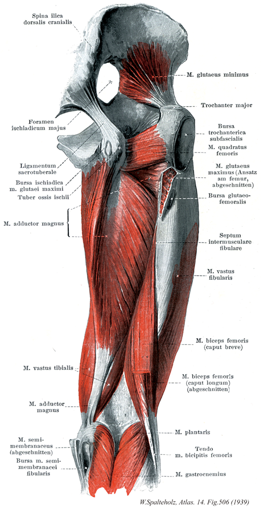
- 506_01【Anterior superior iliac spine; Iliospinale anterius上前腸骨棘;前腸骨棘 Spina iliaca anterior superior; Spina ilica ventralis】 Bony projection at the anterior border of the iliac crest giving origin to the sartorius muscle.
→(腸骨稜の前端は鈍円な突起として大きく突出し、上前腸骨棘として体表上からもよく触れる。大腿筋膜張筋および縫工筋が起こる。)
- 506_02【Greater sciatic foramen大坐骨孔 Foramen ischiadicum majus】 Foramen that is formed by the greater sciatic notch, the sacrospinous ligament, and the sacrotuberous ligament. The piriformis muscle passes from the pelvis through the foramen, producing the following two crevicelike openings.
→(寛骨の大坐骨切痕は、仙結節靱帯と仙棘靱帯によって下方を閉ざされて大坐骨孔になる。この孔は梨状筋が通ることによって、さらに梨状筋上孔と梨状筋下孔とに分かれ、前者を上臀動静脈・神経が通り、後者を坐骨神経のほか下臀動静脈・神経、内陰部動静脈、陰部神経、後大腿皮神経が通る。)
- 506_03【Sacrotuberous ligament; Sacrotuberal ligament仙結節靱帯 Ligamentum sacrotuberale; Ligamentum sacrotuberosum】 Strong band that extends from the sacrum and the ilium to the ischial tuberosity.
→(仙結節靱帯は三角形をした強大な靱帯で、坐骨結節よりおこり、内上方に扇形に放散して、下後腸骨棘、仙骨下半部の外側縁、鼻骨につく。仙棘靱帯とともに、大坐骨切痕および小坐骨切痕をそれぞれ大坐骨孔、小坐骨孔にかえる。また後面は大臀筋の起始となる。しばしば下臀皮神経の枝によって貫かれる。この靱帯の深層で、これと仙棘靱帯との間を、陰部神経、内陰部動静脈が走る。)
- 506_04【Sciatic bursa of gluteus maximus大殿筋の坐骨包;大臀筋の坐骨包 Bursa ischiadica musculi glutei maximi】 Bursa situated between the ischial tuberosity and the inferior surface of the gluteus maximus.
→(大臀筋の坐骨包は大臀筋の深層面と坐骨結節との間にある滑液包で、しばしば欠如する。)
- 506_05【Ischial tuberosity坐骨結節 Tuber ischiadicum】 Bony prominence at the inferior border of the lesser sciatic notch.
→(小坐骨切痕より下方の坐骨体は、その後面に大きな楕円形の坐骨結節を作っている。坐骨結節の表面は大腿後面の筋群に起始を与えるために非常に粗になっている。また坐骨結節は腰掛けるときに椅子の面に接して、体重を支える場所である。)
- 506_06【Adductor magnus muscle大内転筋 Musculus adductor magnus】 o: Inferior pubic ramus, ramus of ischium. i: Medial lip of linea aspera and, via a long tendon, the medial epicondyle. Adduction, lateral rotation, and extension at the hip joint. I: Obturator and tibial nerves.
→(大内転筋は内転筋群の中で最強、人体中最大の筋の1つである。他の内転筋の背側、恥骨下枝および隣接する坐骨枝から坐骨結節までを起始とする。他の起始をもつ部分はほとんどが粗線内側唇に筋性停止する。)
- 506_07【Vastus medialis muscle内側広筋;脛側広筋 Musculus vastus medialis; Musculus vastus tibialis】 o: Medial lip of linea aspera.
→(内側広筋は粗線内側唇の近位2/3と長および大内転筋停止腱から起こる。近位の線維は斜めに下行し、遠位の線維はほとんど横走する大腿四頭筋の共通停止腱に加えて内側広筋の線維は膝蓋骨内側縁や内側膝蓋支帯へ達する。)
- 506_08【Semimembranosus muscle半膜様筋 Musculus semimembranosus】 o: Ischial tuberosity. i: Medial condyle of tibia and oblique popliteal ligament. It is partly covered by the semitendinosus muscle. Extension at the hip joint; flexion and medial rotation at the knee joint. Tenses the knee joint capsule. I: Tibial nerve.
→(半膜様筋は大腿二頭筋長頭と大腿方形筋の起始の間の坐骨結節から起こる。脛骨内側顆、膝関節包後壁および膝窩筋の筋膜に停止する。半膜様筋は中4分の2のみが筋性である。起始腱は広い腱性の板をなし、停止腱も同じ平板である。3本の腱様の索として終わる。脛骨への索は腹側で迂回し、内側側副靱帯の下の脛骨内側顆に付く。中央の索は筋の方向を受け継ぎ、一部は脛骨近位端後面に、一部は膝窩筋の筋膜に付く。腓骨への索は膝関節包の後壁を補強し、斜膝窩靱帯として大腿骨外側顆に向かって外側へ射創する滑液包が通常同筋の停止腱と脛骨内側顆の間にある。)
- 506_09Brodie's bursa【Semimembranosus bursa半膜様筋の滑液包;半膜様筋包 Bursa musculi semimembranosi】 Bursa situated between the tendon of insertion of the semimembranosus and the superior border of the tibia.
→(半膜様筋の滑液包は半膜様筋の停止腱と腓腹筋内側頭との間、および前者と脛骨上端部内側面との間に存在する。)
- 506_10【Gluteus minimus muscle小殿筋;小臀筋 Musculus gluteus minimus】 o:Ilium between the anterior and inferior gluteal lines, i: Greater trochanter. Abduction, medial, and lateral rotation, flexion, and extension at the hip joint. I: Superior gluteal nerve.
→(小臀筋は前および下臀筋線の間の腸骨外側面に起こる。大転子前面の外側縁に停止する。小臀筋はほとんど完全に中臀筋におおわれている。両筋はその前縁で癒合し、後方に開く袋を形成する。小臀筋の転子包は大転子先端と同筋停止腱の間にある。中臀筋と小臀筋は同じ筋原基から生じる。両筋は、大臀筋に対して、小さな臀筋群を作る。大腿屈曲時以外では、股関節を外転するので、外転筋群とすることができる。しかし、大腿に対する作用よりも重大なのは歩行あるいは片足立ちの際に骨盤に対する支持脚の作用である。中小両臀筋は骨盤が遊脚側に傾くのを防ぐ。また、骨盤を支持脚側へ傾ける。)
- 506_11【Greater trochanter of femur大転子(大腿骨の) Trochanter major】 Large prominence on the superolateral aspect of the femur for attachment of the gluteus medius, gluteus minimus, and piriformis muscles.
→(大腿骨頚の上外側には大転子(中臀筋、小臀筋、梨状筋がつく)が突出している。転子とはハンドルのことで、その力学的な効用は、たとえば大転子につく中臀筋が大腿骨を外転させている。)
- 506_12【Subcutaneous trochanteric bursa皮下転子包;大転子皮下包 Bursa subcutanea traochanterica】 Bursa situated on the tendon of the gluteus maximus between the skin and the greater trochanter.
→(皮下転子包は大臀筋の表層で大転子部の皮下にある。)
- 506_13【Quadratus femoris muscle大腿方形筋 Musculus quadratus femoris】 o: Ischial tuberosity. i: Intertrochanteric crest. Lateral rotation and adduction. I: Sacral plexus.
→(大腿方形筋はほとんど筋性で、大腿骨が正常位にあれば四辺形である。坐骨結節から転子間稜へ横走する。筋の大きさから予想する以上にこの筋が効果的に股関節外施に働くのは筋力のほとんどが外施に有効となるような筋線維走行だからである。)
- 506_14【Gluteus maximus muscle大殿筋;大臀筋 Musculus gluteus maximus】 o: Ilium, behind the posterior gluteal line, sacrum, coccyx, thoracolumbar fascia, sacrotuberous ligament, i: Fascia lata, iliotibial tract, gluteal tuberosity, lateral femoral intermuscular septum, linea aspera. Extension, lateral rotation, abduction, and adduction at the hip joint. I: Inferior gluteal nerve.
→(大臀筋は大腿を伸展する主力筋で、とくに歩行の際重要である。中臀筋や小臀筋(小さな臀部の筋群)と同様、大きな臀部の筋である大臀筋も発生的には伸筋群である。仙骨と尾骨の辺縁、後臀筋線より後方の腸骨稜、胸腰筋膜、そして仙結節靭帯などから起始する。その厚い筋線維束は斜め下方へ走り、広い停止腱となる。その停止域は近位では大腿筋膜、腸脛靱帯に放散する。また、臀筋粗面よりも遠位で外側筋間中隔より上の粗線外側唇にも停止する。坐骨包坐骨結節と大臀筋下面の筋膜との間にある。慢性的刺激の結果として(機織工結節、抗夫結節)、臀部に敷物なしに座り仕事をする人々では同包に炎症が起こり、後大腿皮神経を圧迫する。大腿筋の停止腱は転子包によって大転子と離される。臀筋粗面では、大腿筋はふつう他の臀筋との間にあるいくつかの筋間包の上を滑走する。立位では大臀筋下部が坐骨結節をおおう。大腿を屈すると大臀筋下部は頭側に移動する。このため座位では坐骨結節は皮下脂肪上に位置し、皮膚を通して容易に触れる。臀溝はほぼ水平に走り、大臀筋収縮時には深くなるが、大臀筋の下縁をあらわしているわけではなく、同筋走行に対して鋭角的に交わる。)
- 506_15【Intermuscular gluteal bursae筋間包(殿筋の);殿筋筋間包;大臀筋大腿骨包 Bursae intermusculares musculorum gluteorum; Bursae glutaeofemorales】 Two or three bursae situated beneath the insertion of the gluteus maximus on the gluteal tuberosity.
→(臀筋の筋間包は大臀筋の腸脛靱帯移行する停止腱膜の深層で、これと外側広筋との間にある。)
- 506_16【Lateral femoral intermuscular septum外側大腿筋間中隔 Septum intermusculare femoris laterale; Septum intermusculare fibulare】 Strong layer of connective tissue formed by the fascia lata at the lateral lip of the linea aspera between the biceps femoris and vastus lateralis.
→(外側大腿筋間中隔は大腿筋膜の一部が大腿骨粗線の外側唇に付着したもので、外側広筋と大腿二頭筋短頭の間に張り、両筋の一部筋束はこれより起始する。大腿骨における大臀筋の停止部から大腿筋外側顆の間にのびる。)
- 506_17【Vastus lateralis muscle外側広筋;腓側広筋 Musculus vastus lateralis; Musculus vastus fibularis】 o: Greater trochanter, lateral lip of linea aspera.
→(外側広筋は4頭のうち最大で、大転子基部、粗線外側唇および大転子から発する表在性腱膜から起こる。膝蓋骨よりも近位で腱となり、大腿四頭筋の共通腱に合流する。また、一部の腱線維は膝蓋支帯へ放散する。)
- 506_18【Biceps femoris muscle大腿二頭筋 Musculus biceps femoris】 Two-headed muscle arising from the pelvis and femur, i: Head of fibula. Flexion at the knee joint, lateral rotation.
→(大腿二頭筋は2関節性の長頭と1関節性の短頭から成る。長頭は坐骨結節で半腱様筋と総頭をつくって起こる。短頭は粗線の外側唇の中1/3と外側筋間中隔から起こる。これら両頭は合して2頭筋となり、腓骨頭に終わる。その際この筋と膝関節の外側側副靱帯との間に大腿二頭筋の下腱下包がある。股関節では長頭は大腿を後斜するように働く。膝関節では大腿二頭筋は屈曲するように働き、屈曲した状態では下腿を外旋する。この筋は膝関節における唯一の外旋筋であって、すべての内旋筋に匹敵する作用をもっている。)
- 506_19【Short head of biceps femoris muscle短頭(大腿二頭筋の) Caput breve (Musculus biceps femoris)】 o: Lateral lip of linea aspera. I: Common fibular nerve.
→(大腿二頭筋の短頭は粗線の外側唇より起こり、腓骨頭に終わる。腓骨神経より支配される。作用として膝関節の屈曲および外旋。)
- 506_20【Long head of biceps femoris muscle長頭(大腿二頭筋の) Caput longum (Musculus biceps femoris)】 o: Ischial tuberosity. Extension at the hip joint. I: Tibial nerve.
→(大腿二頭筋の長頭は半腱様筋とともに坐骨結節後面から起こる。短頭は粗線外側唇の中1/3から起こる。同レベルの粗線内側唇に停止する長内転筋の線維方向を短頭は引き継ぐ。両頭の共通停止腱は腓骨頭に止まる。腱線維の一部は脛骨外側顆および下腿筋膜に達する。大腿二頭筋腱は腱下包によって外側側副靱帯と隔てられる。)
- 506_21【Plantaris muscle足底筋 Musculus plantaris; Musculus plantaris longus】 o: Above the lateral femoral condyle. i: Achilles tendon or calcaneus tuberosity. I: Tibial nerve.
→(足底筋は腓腹筋とヒラメ筋の間にあり、結合組織に囲まれている筋。腓腹筋外側頭の内側、大腿骨外側顆の外側上顆稜から起こる。細い筋腹から細く長い停止腱が腓腹筋とヒラメ筋の間を走り、(アキレス腱を介して)踵骨隆起へ至る。同筋はときどき下腿筋膜に、あるいはまれには足底腱膜に続く。足底筋は下等霊長類にみられる足底腱膜へ続く筋の、系統発生的な、遺残筋である。人ではときどき欠如したり腓腹筋外側頭と癒合したりする。足底筋は腓腹筋とヒラメ筋の間にあって、結合組織に包まれている。その結合組織は後脛骨動静脈の血管鞘と連絡している。同筋はヒラメ筋腱弓よりも上ではこれらの血管を守るようにその上を走る。この筋は脛骨神経支配を受け、距腿関節による足の底屈、膝関節の屈曲を助けるが、その力は弱い。膝関節屈曲中に足底筋は神経血管索を引き上げ、弓状走行をとらせ、血管が折れ曲がるのを防ぐ役割をする。)
- 506_22【Biceps femoris tendon大腿二頭筋腱 Tendo musculus biceps femoris】
→()
- 506_23【Gastrocnemius muscle腓腹筋 Musculus gastrocnemius】 Superficial leg muscle composed of the following two heads. Flexion at the knee joint, plantar flexion and supination at the ankle joint.
→(下腿後側の浅層の筋で、ヒラメ筋と合わせて下腿三頭筋と呼ばれる。大腿骨の内側上顆(内側頭)と外側上顆(外側頭)とから起こる。その停止腱はヒラメ筋とともに合流し、アキレス腱(踵骨腱)となり、踵骨隆起に停止する。踵骨後面の上部と踵骨腱が接近する部位には小さな滑液包が介在する。この筋は脛骨神経支配を受け、距腿関節による足の底屈、膝関節屈曲を生じさせる。Gastrocnemiusはフクラハギ(gastro-腹[腹のようにふくらむ]+kneme脚、スネ))