Spalteholz HANDATLAS DER ANATOMIE DES MENSCHEN VON WERNER SPALTEHOLZ
メニューは解剖学(TA)にリンクしてあります。図の番号をクリックすると下記の説明へ、右側の用語をクリックすると解剖学(TA)にジャンプします。
531 


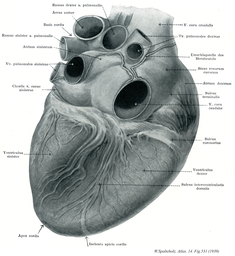
- 531_00【Heart心臓 Cor】
→(心臓は血液循環の原動力となる器官で静脈から血液を受け取り、動脈に送り出す中空の筋肉の器官。その壁は主として心筋組織でできている。心臓は心膜に包まれて、縦隔の前下部で左右の肺の間に置かれ横隔膜の上面にのる。全形を円錐にみたてると、その軸は後上右方(心尖)へ向かう。心臓の約3分2が正中線の左方、3文の1が右方にある。心尖(左心室の尖端)の拍動は第5肋間隙で左乳頭線のやや内側に触れる。心臓の内部は4部屋に分かれ(右と左の心房・心室)、心房中隔および心室中隔が左右を隔てる。各側の心房と心室は房室口を介して連なる。外面では、冠状溝が心房と心室の境をかこみ、前・後の室間孔は両親質の境をあらわす。これらの溝を冠状血管の主枝が走る。左右の心事は各心房の一部が前方に突出したもので、動脈管のねもとを両側から抱く形である。心耳の壁はうすく内面に櫛状筋が発達している。左心耳は右心耳に比べて格段に小さい。右心房には上大静脈・下大静脈そして冠状静脈洞が開く。前二者が開口する部域はもと静脈洞の右角に由来し内面が平滑である(大静脈洞)。この部域と右心耳との境目に分界稜が隆まる。これに体操して外側には分界溝があらわれる。心房中隔の右側面には卵円窩が卵円孔の閉じた跡を示す。左側面では前方よりに中隔鎌とよぶヒダ(卵円孔弁のなごり)がみえる。左心房は心臓の最背側に位置を占め、左右から各2本の肺静脈が ここに注ぐ。[心臓の弁]左・右心室の出入口それぞれ弁装置をそなえ、そのしくみにより血液が逆流するのをふせいでいる。右房室口には3枚の帆状弁があり三尖弁(右房室弁)、左房室口2枚あり僧帽弁(左房室弁)とよぶ。そして各心室の内面に乳頭筋という指状の高まり(右室では3群、左室では2群)があって、それが弁膜の縁と複数の腱索で結ばれている。心室が収縮するとき帆状弁は翻転することなく互いに接しあい房室口を閉じる仕組みである。心室の出口側は、肺動脈口(右心室より肺循環へ)および大動脈口(左心室より体循環へ)にそれぞれ3個のポケット形の半月弁がある。各弁膜の自由縁は中央が肥厚し(半月弁結節)、この両脇が薄くできている(半月)。3個のポケットが接し合うことによって動脈口は閉じる。このしくみは乳頭筋とは関係がない。心室中隔は大部分が筋性であるが、ただ一部(大動脈口の直下)に筋質を欠き薄くできた場所がある(膜性部)。これにつづき左心室と右心房とを隔てる膜性の構造があり、それを房室中隔とよぶ。左心室はビヤ樽形でその壁の厚さは右心室の側壁の約3倍。心室中隔は右室腔に向かってまるく凸面をなす。右心室の内腔はV字形であり、流入部と流出部とに分かれ、筋質の室上稜が両者を境する。右心室の流出部を動脈円錐ともよぶ。心臓は左側にあると思って思っている人が多いが、これは心尖拍動が左に触れられることから生じた誤解である。心臓は多少左にずれているとはいえ、縦隔の中部でほぼ正中に位置している。心臓の大きさは、生体では心室の収縮期systoleと拡張期diastoleで異なる。また心臓の位置も、呼吸運動に伴って横隔膜が上下するので、生体では絶えず移動しているわけである。深く息を吸い込むと、心臓は左右の肺に圧迫されるばかりでなく、横隔膜が下に下がるので、多少縦長になる。逆に息を深く吐いたときには、心臓はずんぐり形になる。心臓の大体の大きさとか位置や形は生体でも打診percussionによって知ることができ、その変化が色々な病気の診断の一つの手掛かりになる。)
- 531_10【Superior vena cava上大静脈 Vena cava superior; Vena cava cranialis】
→(上大静脈は上半身の血液を集める静脈で、上縦隔の中で左右の腕頭静脈が合してはじまり、途中で奇静脈を受け入れながら上行大動脈の右側を下行して右心房にそそぐ。)
- 531_13【Right atrium右心房 Atrium cordis dextrum; Atrium dextrum】
→(右心房は心臓の右上部を占め、その後上部と後下部とに、それぞれ、上大静脈と下大静脈が注いでいる。)
- 531_16【Coronary sulcus冠状溝 Sulcus coronarius】 Groove that runs around the heart, demarcating the borders between the atria and ventricles.
→(心房と心室の境には冠状溝とう溝があるが、冠状溝は冠状動脈と冠状静脈洞で埋められている。)
- 531_17【Right ventricle右心室 Ventriculus dexter】
→(右心室は心臓の最下位部を占め、後上方にある右房室口で右心房と交通し、前上方にある肺動脈口で肺静脈に連なる。)
- 531_19【Notch of cardiac apex; Apical notch of heart心尖切痕 Incisura apicis cordis】 Indentation to the right of the apex where the two interventricular sulci become continuous with each other.
→(心尖切痕は室間溝の延長にある、心尖近く右側の切れ込み。)
- 531_02【Arch of aorta; Aortic arch大動脈弓 Arcus aortae】 It is located between the ascending and descending aorta. Its roof extends to the first rib at the left border of the sternum.
→(大動脈弓は上行大動脈につづく弯曲部であり(約5~6cm長)、肺動脈分岐部および左気管支をこえて左後方にまわり、第四胸椎体の左側で胸大動脈に移行する。)
- 531_05【Left atrium左心房 Atrium cordis sinistrum; Atrium sinistrum】
→(左心房は心臓の後上部にあって、後面をつくっている。左心房は右心房よりもやや小さいが、壁はやや厚い。左心房の後壁の上部に、左右両肺からそれぞれ2本ずつ、前部で4本の肺静脈が開口している。左心房は前下方で房室口によって左心室に通じる。)
- 531_08【Left ventricle左心室 Ventriculus sinister】
→(左心室は心臓の左下部を占め、後上方にある左房室口で左心房と交通し、右上隅にある大動脈口によって大動脈につらなる。左心室の壁は右心室に比べ2~3倍厚い。心室中隔は、右心室に向かって膨隆しているので、心室を横断面でみると、左心室の内腔は円いのに対して、右心室の内腔は半月状である)
- 531_09【Apex of heart心尖 Apex cordis】 Part of the heart directed downward and to the left. It is formed by the left ventricle.
→(心尖は心臓の下端部でやや尖っている。心尖は心臓の拍動とともに前胸壁にあたる。これを心尖拍動といい、体表で触れることができる。すなわち、一般に左側の第5肋間で、正中線から約4横指(約7cm)左方で触れる。この位置は弾性では左乳頭のすこし内下方である。小児ではやや高くかつ外方にある。)
- 531_00a【Diaphragmatic surface of heart; Inferior surface of heart横隔面;下面(心臓の) Facies diaphragmatica cordis; Facies inferior cordis】 Inferior, flattened surface of the heart that is in contact with the diaphragm.
→(心臓の横隔面は横隔膜に接する心臓の下面で、主として横隔膜の腱中心の上にある。ほぼ平坦で、心室とくに左心室と右心室の一部でできる。)
- 531_01【Right pulmonary artery右肺動脈;右枝(肺動脈の) Arteria pulmonalis dextra; Ramus dexter】 Artery located behind the ascending aorta. Its ramifications parallel the branches of the bronchial tree. Both form bronchoarterial segments.
→(右肺動脈は肺動脈間からの2分枝のうちの長い方で縦隔上部で正中線を横切って右肺門に肺根の一部となって加わる。その分枝は気管支や細気管支に伴行して分布するが個人差が大きい。典型的には①上葉動脈から肺尖動脈(A1)と前上葉動脈(A3)・後上葉動脈(A2)、②中葉動脈から外側枝(A4)、内側枝(A5)③下葉動脈から上区動脈が出る。④下葉動脈の肺底部からは前肺底動脈、後肺底動脈、外側肺底動脈、内側肺底動脈がでる。)
- 531_03【Base of heart心底 Basis cordis】 Broad aspect of the heart facing dorsally and to the right, located opposite to the apex of the nearly conical heart, it is mainly formed by the posterior wall of the left atrium. The pulmonary arteries and vasa privata arise and open here.
→(心底はほぼ円錐状の心臓の上側、心尖の反対側の広い面。主に左心房と右心房の一部とできる。横隔面からは冠状溝で境される。)
- 531_04【Left pulmonary artery左肺動脈;左枝(肺動脈の) Arteria pulmonalis sinistra; Ramus sinister】 Artery lying in front of the descending aorta. On radiographs it appears as a 「pulmonary arch」 below the 「aortic arch.」
→(左肺動脈は肺動脈幹の2分枝のうち短い方の枝で、心外膜を貫いて左肺門にはいる。多数の枝を出して気管支区や気管支下区に分布するが個人差は大である。典型的には上葉動脈の枝として肺尖動脈(A1)、前区動脈、後区動脈(後2者は上行枝・下行枝を出す)、肺舌動脈の枝として上舌動脈・下舌動脈、下葉動脈の枝として下葉上動脈(A6)と肺底区の枝(前肺底動脈、後肺底動脈、外側肺底動脈、内側肺底動脈)を出す。)
- 531_06【Left pulmonary veins左肺静脈 Venae pulmonales sinistrae】 The two left pulmonary veins which occasionally unite to form a single trunk.
→(2条。ときには合して1本の幹となる。 (Feneis))
- 531_06a【Left superior pulmonary vein左上肺静脈;上左肺静脈 Vena pulmonalis sinistra superior】 Pulmonary vein that drains the left superior lobe.
→(左上肺静脈は左上葉から血液を運び、左主気管支より腹側やや尾側を、そして胸大動脈の前方を通る。左上肺静脈は肺尖後静脈、前上葉静脈、肺舌静脈が合流して形成される。)
- 531_06a【Pulmonary veins肺静脈 Venae pulmonales】 Blood vessels leading from the lungs to the heart.
→(肺静脈は正常では肺から上肺静脈と下肺静脈が、酸素に富む血管を肺胞壁の毛細血管網から左心房に運ぶ。また肺組織や細いまたは中等大の気管支の壁からも血液を受ける。両側の肺門では、肺静脈は肺門の前下縁近くに位置している。上・下肺静脈は左心房に開口する前に共通幹を形成することがある。)
- 531_06b【Left inferior pulmonary vein左下肺静脈;下左肺静脈 Vena pulmonalis sinistra inferior】 Pulmonary vein that drains the left inferior lobe.
→(左下肺静脈は、右下肺静脈と同じ名称の「幹」静脈からなり、気管支の前で外側肺底区、後肺底区から左心房へ至る。)
- 531_07【Ligament of left vena cava左大静脈靱帯;左上大静脈靱帯;左大静脈ヒダ Ligamentum venae cavae sinistrae; Chorda venae cavae sinistrae】 Pericardial fold formed by the connective-tissue cord of the obliterated embryonic left superior vena cava. It lies in front of the left pulmonary vessels, which it ca. connect with one another.
→(左大静脈ヒダは腕頭静脈と斜静脈(胎生時の左上大静脈遺残)の間の結合組織索により隆起した心膜のヒダ。左肺血管の前方に位置し、相互に結合しうる。)
- 531_11【Right pulmonary veins右肺静脈 Venae pulmonales dextrae】 The two right pulmonary veins which occasionally unite to form a single trunk.
→(2本あるが、時に合流して1本の幹となる。 (Feneis))
- 531_11a【Right superior pulmonary vein右上肺静脈;上右肺静脈 Vena pulmonalis dextra superior】 Pulmonary vein that drains the superior and middle lobes.
→(右上肺静脈は、短い静脈幹すなわち肺尖静脈、前上葉静脈、後上葉静脈を介して上葉の区域から、また中葉静脈を介して中葉から血液を集める。そして右肺静脈より腹尾側、かつ上大静脈の後方で左心房に到達する。)
- 531_11b【Right inferior pulmonary vein右下肺静脈;下右肺静脈 Vena pulmonalis dextra inferior; Vena pulmonalis inferior dextra】 Pulmonary vein that drains the right inferior lobe.
→(右肺下葉から左心房に酸素化された血液を戻す静脈。右下葉からの上肺底静脈や総肺底静脈が流入する。)
- 531_12【Sinus of venae cavae大静脈洞 Sinus venarum cavarum】 Smooth-walled space bounded by the crista terminalis for the passage of blood from the inferior and superior venae cavae.
→(両大静脈から血液を受け入れる腔で、平滑な壁をもち分界稜で境される。 (Feneis))
- 531_14【Sulcus terminalis cordis; Terminal sulcus of right atrium分界溝;右心房分界溝 Sulcus terminalis cordis; Sulcus terminalis atrii dextri】 Groove visible on the external surface of the right atrium between the embryonic sinus venosus and atrium. It surrounds the region surrounding the openings of the inferior and superior venae cavae.
→(発生学的に静脈洞と固有心房の間の境にある、外から見える溝。上下大静脈の開口部領域を取り囲んでいる。)
- 531_15【Inferior vena cava下大静脈 Vena cava inferior; Vena cava caudalis】 It arises at the union of the right and left common iliac veins, lies on the right side of the aorta, and opens into the right atrium of the heart.
→(下大静脈は下肢および骨盤と腹部の器官の大部分から血液を受ける本幹で、第5腰椎体の右側で左右の総腸骨静脈の合流として始まり、このあと脊柱に沿って大動脈の右側を上行、肝臓の後面をこれに接して通過し、第八胸椎の高さで横隔膜の大静脈孔を貫いて胸腔に入り、ただちに右心房にそそぐ。下大静脈に流入する枝には総腸骨静脈、下横隔静脈、第3・第4腰静脈、肝静脈、腎静脈、右副腎静脈、右精巣静脈、右卵巣静脈、蔓状静脈叢などがある)
- 531_18【Posterior interventricular sulcus後室間溝 Sulcus interventricularis posterior; Sulcus interventricularis dorsalis】 Longitudinal groove on the diaphragmatic surface of the heart corresponding to the interventricular septum. It transmits the posterior branch of the interventricular branch of the right coronary artery.
→(後室間溝は横隔面で心室中隔に層とする縦溝。右冠状動脈の後室間枝が走る。)