Spalteholz HANDATLAS DER ANATOMIE DES MENSCHEN VON WERNER SPALTEHOLZ
メニューは解剖学(TA)にリンクしてあります。図の番号をクリックすると下記の説明へ、右側の用語をクリックすると解剖学(TA)にジャンプします。
983


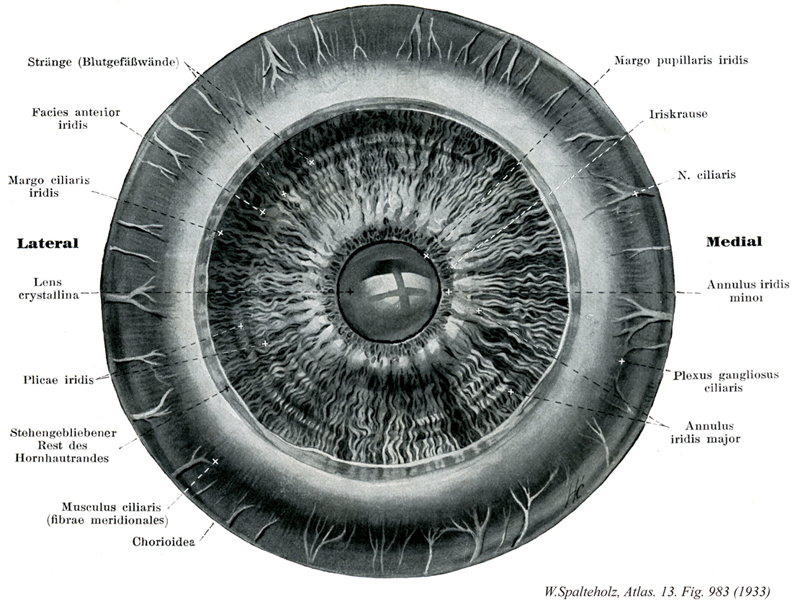
- 983_00【Ciliary body毛様体 Corpus ciliare】 Thickened area situated between the ora serrata and root of the iris that contains the ciliary muscle and ciliary processes.
→(毛様体は虹彩支質と脈絡膜の移行部に生後発生する平滑筋(毛様体筋)の束により生じる肥厚部である。経線断面では全体として三角形の断面をもち、3頂点がそれぞれ網膜鋸状縁、虹彩基部、毛様体突起にあたる。毛様体突起には毛様体小帯が結合し、水晶体に連絡する。眼球を赤道面で切断し、その前半分を後ろから観察すると水晶体を一周する毛様体を一眺することができる。鋸状縁につづく、①毛様体輪(幅~3mm)と、②放射状に配列する約70個の毛様体突起とヒダよりなる毛様体間が区別される。輪部には細い経線方向に走る稜線が認められ、これが集中して毛様体突起(長さ2mm、幅1mm、高さ0.5mm)をつくる。)
- 983_01【Anterior surface of iris前面(虹彩の) Facies anterior iridis】 Anterior surface of the iris facing the anterior chamber.
→(前眼房へ対する面。(Feneis))
- 983_02【Ciliary margin of iris毛様体縁(虹彩の) Margo ciliaris iridis】 Outer border of the iris that is attached to the ciliary body and at the iridocorneal angle.
→(虹彩の毛様体縁は虹彩の外縁。毛様体および虹彩角膜角へ固定される。)
- 983_03【Lens水晶体 Lens; Lens crystallina】 Lens suspended by the ciliary zonule between the pupil and vitreous body. It measures 9-10 mm in diameter and is about 4 mm thick.
→(水晶体は虹彩の後方、硝子体の前方に位置し、双凸面レンズ構造をもつ。赤道直径約9mm、水晶体軸(前、後極を結ぶ直線)3.7~4.4mm、前面弯曲度から8mm、後面弯曲度~6mm、屈折率1.36(中央部)~1.42(辺縁部)。水晶体は無色透明な水晶体包(前面で厚く、後面で薄い粘液多糖体層で、水晶体上皮の基底膜が発達したもの)におおわれる水晶体質よりなる。水晶体質はより軟らかい上皮と硬い核に分かれやすく、胎児では雌で水晶体包に切れ目をいれるとはじけるように裂ける。生体では前、後極から発する数本の水晶体放線がわずかに認められ、胎児では前後両面放線がわずかに認められ、胎児では前後両面に、たがいに120°に交わる3本の放線(前面逆Y字、後面正Y字形)を示す。水晶体の構成要素は水晶体線維で、発生初期の単層の水晶体胞の後壁の細胞のみが著しく長大化したものである。前面に層単層の水晶体上皮は水晶体胞前壁の原型を保つ。赤道より後面にいくにしたがい長細い六角形の水晶体線維の束へと移行する。胎児期の放線は水晶体線維束の付着点をなす中隔に一致し、前極からおこる線維は後面の赤道近くの最寄りの放線に、前面赤道近くの中隔よりおこる線維は後極へ向かう。水晶体線維は緊密かつ生前と配列するが、微絨毛を出して細い細胞管腔を確保し、水および代謝物質の移送路を形成する。生体の水晶体には血管の神経の分布が認められない。胎児の水晶体包は硝子体動脈により養われるが、妊娠末期に道動脈が閉鎖する。老年者では前後面の弯曲度が減って扁平となり、黄白色を帯びる傾向にある。この変化が進行したものを白内障cataractという。全体の25%を占める水晶体蛋白はα-およびβ-クリスタリンと不溶性アルブモイドよりなり、そのほかにグルタチン、ビタミンCなどが含まれる。)
- 983_04【Folds of iris虹彩ヒダ Plicae iridis; Rugae iridis】 Folds passing around the border of the anterior surface of iris. They produce the slightly scalloped appearance of the border of the iris.
→(虹彩前面の瞳孔縁周囲にあるヒダ。瞳孔縁に浅い鋸歯状構造をつくる。(Feneis))
- 983_05【Corneoscleral junction; Corneal limbus角膜縁 Limbus corneae; Margo corneae】 Margin of the cornea that is continuous with the sclera.
→(角膜縁は角膜の強膜への移行縁で強膜のへこんだ境界縁。)
- 983_06【Ciliary muscle毛様体筋 Musculus ciliaris】 Smooth muscle of the ciliary body. It draws the choroid anteriorly, thereby relaxing the zonular fibers, allowing the lens to take its own, more strongly convex form for near vision.
→(眼の遠近調節accommodation(ピント合わせ)はの仕組みは、近いものを見るときには、毛様体筋が収縮して毛様小体がゆるみ(毛様小体の付着部が前の方に引かれるため)、水晶体が自身の弾性によって膨らみ(球形に近付き)、屈折率を増す。長時間にわたって細かい字を読んだ時の眼の疲労は、主に毛様体筋の疲労である。)
- 983_07Bücke's muscle【Meridional fibres of ciliary muscle経線状線維;経線線維(毛様体筋の);ブリュッケの筋 Fibrae meridionales (Musculus ciliaris)】 Larger group of fibers running meridionally that mostly arise from the scleral spur and pass along the choroid to the sclera.
→(ブリュッケ筋とも呼ばれる。毛様体筋のうちの経線状に走る平滑筋線維。筋の大部分を占める。前方、後境界板のところは虹彩角膜角櫛状靱帯へ、後方は脈絡膜へつく。眼球の毛様体内部に含まれる。オーストラリアの生理学者Ernest W. von Bruecke (1819-1892)の名を冠する。この筋はアイルランドの外科医Sir Philip Crampton (1777-1892)の名を冠する。この筋はアイルランドの外科医Sir Philip Crampton (1777-1858)のなをとってCrampton's muscleとも呼ばれる。なお、毛様体の輪状平滑筋はミュラー筋(Mueller's muscle)と呼ばれている。)
- 983_08【Choroid脈絡膜 Choroidea; Chorioidea】 Portion situated between the retina and sclera.
→(脈絡膜は網膜視部の外側に接してこれを包んでおり、内側から外側に向かって、①基底膜、②脈絡毛細血管板、③血管板、④脈絡上板の4層に分けられる。)
- 983_09【Pupillary margin of iris瞳孔縁(虹彩の) Margo pupillaris iridis】 Inner border of the iris encircling the pupil.
→(虹彩の瞳孔を囲む縁。(Feneis))
- 983_10【Iris虹彩 Iris】 Round disc with a central opening (pupil) situated in the frontal plane that varies in color in different individuals. It forms the posterior end of the anterior chamber and becomes continuous at its margin with the ciliary body. It has a diameter of 10-12 mm.
→(虹彩は、前頭面に位置し、眼の血管層の前方部分をつくる隔膜で、色に個人差のある円板。中央に開口部(瞳孔)があり、直径は約10~12mm。前眼房の後境界で、その縁は毛様体へ移行する。周囲辺縁は強膜岬角に付着している。瞳孔をかたちづくるあたかもカメラの絞りのような器官で、虹彩内皮、虹彩支質、虹彩筋、虹彩色素上皮層より構成され、血管に富む。虹彩の脈管と神経はは虹彩の動脈としては毛様体縁に沿う大虹彩動脈輪、瞳孔縁に沿う大虹彩動脈輪、瞳孔縁に沿う小虹彩動脈輪、両者を放射状につなぐ小動脈があり、長後毛様体動脈、前毛様体動脈、脈絡膜毛細血管叢より供給される。静脈血はこれらに伴う静脈のほか、渦静脈に流入する。虹彩の神経支配として長毛様体神経(三叉神経由来の体知覚性神経)と短毛様体神経(毛様体神経節由来の自律神経)があり、後者には動眼神経副核由来の節前線維から興奮を受けて伝達する節細胞の軸索すなわち副交感神経節後線維と、内頚動脈神経叢を経て毛様体神経節に達し、節内でそれに合流する胸部交感神経核由来の交感神経節後線維が含まれる。瞳孔括約筋は副交感神経、散大筋は交感神経の支配を受ける。)
- 983_11【Ciliary nerves毛様体神経 Nervi ciliares】
→()
- 983_11a【Long ciliary nerves長毛様体神経 Nervi ciliares longi】 Two, long, thin branches that carry sympathetic fibers to the dilator pupillae and afferent fibers from the iris, ciliary body, and cornea.
→(通常2本あって、毛様体神経節から出る短毛様体神経とともに視神経の付近で眼球に入り、強膜を貫いて、これと脈絡膜の間を前にすすみ、毛様体と虹彩にいたる鈍知覚枝である。)
- 983_11b【Short ciliary nerves短毛様体神経 Nervi ciliares breves】 Up to 20 short nerves that pierce the sclera around CN II. They contain postganglionic parasympathetic fibers from the ciliary ganglion as well as postganglionic sympathetic fibers of the sympathetic root that carry information to the eye. They contain sensory fibers of the nasociliary root that carry impulses away from the eye.
→()
- 983_12【Inner border of iris小虹彩輪;内虹彩輪 Anulus iridis minor】 Pupillary portion of the iris. Narrower zone that is distinguishable from the outer border of iris by its more delicate structure.
→(虹彩の瞳孔部。狭く、密な構築で大虹彩輪とは区物される部分。(Feneis))
- 983_13【Ciliary ganglionic plexus毛様体神経節神経叢 Plexus gangliosus ciliaris; Plexus nervosus gangliosus ciliaris】
→()
- 983_14【Outer border of iris大虹彩輪;外虹彩輪 Anulus iridis major】 Ciliary portion of the iris. Outer, wider zone that is distinguishable from the inner border of the iris by its coarser structure.
→(虹彩の毛様体部。外側で、幅広く、粗大な構築で小虹彩輪とは区別される部分。(Feneis))