Spalteholz HANDATLAS DER ANATOMIE DES MENSCHEN VON WERNER SPALTEHOLZ
メニューは解剖学(TA)にリンクしてあります。図の番号をクリックすると下記の説明へ、右側の用語をクリックすると解剖学(TA)にジャンプします。
597 


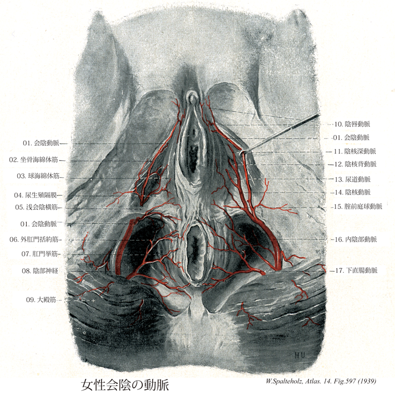
- 597_01【Perineal artery会陰動脈 Arteria perinealis】 Artery arising at the posterior margin of the urogenital diaphragm. It supplies the bulbospongiosus and ischiocavernosus.
→(会陰動脈は下直腸動脈より腹側で本幹より分岐し、浅会陰横筋の上または下を通り、球海綿体筋と坐骨海綿体筋の間を前進して、これらに筋枝を与えたのち、数本の後陰嚢枝(♂)または後陰唇枝(♀)となって、これらの皮膚に分布する。)
- 597_02【Ischiocavernosus muscle坐骨海綿体筋 Musculus ischiocavernosus】 Male: Muscle extending from the ramus of ischium over the cms of the penis to the tunica albuginea. Smaller bundles of muscle fibers run over the penis below the pubic symphysis to the contralateral side.
→(坐骨海綿体筋は男性より女性のほうが発達が弱い。この筋は坐骨枝より起こり陰核脚を被い、その腱性線維は外下表面に付く。その筋は陰核海綿体を圧し、血液を押し付け流出を妨げ、それにより陰核の勃起成立を助ける。)
- 597_03【Bulbospongiosus muscle球海綿体筋 Musculus bulbospongiosus; Musculus bulbocavernosus】 Male: Muscle arising from the perineal body and the inferior aspect of the corpus spongiosum of penis, passing to the perineal membrane and dorsum of penis. It is unpaired. It acts to compress the bulb of penis and transport urethral contents further. ABC Female: Muscle that originates on the ramus of ischium, attaching to and covering the cms of clitoris. It assists in filling the cavernous bodies with blood. I: Pudendal nerve.
→(男性では球海綿体は尿道球の周辺を不体の筋として回るが、会陰の中心腱と尿道海綿体下側の正中縫線から起こる。球海綿体は前方へ放散し、海綿体のまわり下尿生殖隔膜筋膜や尿道海綿体へ向かい、また前筋線維をもって陰茎背部へ付く。この筋は随意的または反射的に尿道球を圧迫し、それにより尿道の内容を駆出する。女性では球海綿体筋は男性のように全長で1つの筋にはなっていない。2つの筋が会陰の中心腱より起こるが、各筋はそれぞれ引き続き前庭球と大前庭腺を被っている。その筋束は前庭球や陰核海綿体に停止し、陰核体後部で反対側からの筋線維と絡み合っている。この筋は大前庭腺を反射的に空にし、血液を前庭球の後方拡大部から送り出し、またオルガスムの際外腟口を収縮させる。)
- 597_04【Urogenital diaphragm尿生殖隔膜 Diaphragma urogenitale】 A term that has been replaced. What was previously conceived of as a unit is now divided into separate terms: perineal membrane, transverse perineal ligament, deep transverse perineal muscle.
→(尿生殖隔膜は骨盤隔膜の前部の下側(浅側)にあり、恥骨弓の間に張っている三角形の線維性膜である。尿生殖膜は上・下2枚の筋膜からなる。この筋膜を、それぞれ、上・下尿生殖隔膜筋膜という。とくに下尿生殖隔膜筋膜は比較的厚く強靱で、会陰膜ともいわれる。上生殖隔膜筋膜は明瞭でないことも多い。上尿生殖隔膜筋膜と下尿生殖隔膜筋膜とは後縁では癒合し、会陰腱中心に付着する。上下の隔膜筋膜は前上縁でも癒合し肥厚して、恥骨結合のすぐ下で会陰横靱帯をつくる。尿生殖隔膜は、男性では尿道によって、女性では尿道と腟とで貫かれる。)
- 597_05Thiele's muscle【Superficial transverse perineal muscle浅会陰横筋 Musculus transversus perinei superficialis】 Inconstant expansion of the deep transverse perineal muscle that extends from the ischial tuberosity to the perineal body. I: Pudendal nerve.
→(浅会陰横筋は横走する浅在性の薄い筋である。この筋は坐骨結節や坐骨枝の境界域の起始部ではしばしば坐骨海綿体筋と交通し、そこから分枝する。その線維は会陰体へ連なり、外肛門括約筋と球海綿体筋に放散する。女性ではその筋は多少退化し、わずかに筋膜の被膜のみ同定できる程度である。)
- 597_06【External anal sphincter muscle外肛門括約筋 Musculus sphincter ani externus】 Muscle with transversely striated fibers.
→(外肛門括約筋は内肛門括約筋の衿のように張り付いている。外口門括約筋のほぼ矢状面に位置する筋束が腸間終端を両側から閉鎖する。この筋束は後方では尾骨から張る靱帯(肛門尾骨靱帯)に付着し、前方では会陰中心に付いている。)
- 597_07【Levator ani muscle肛門挙筋 Musculus levator ani】 Principle muscle of the pelvic diaphragm. It is derived from the abdominal wall musculature and permeated by smooth-muscle cells. I: Sacral plexus, S2-S5. It consists of the following parts.
→(肛門挙筋の丈夫な前部(恥骨尾骨筋)は分界線直下の恥骨の内面から起こり、薄い後部(腸骨尾骨筋)は腸骨から起こる。その起始腱は内閉鎖筋筋膜に接して移行し、閉鎖筋膜から発する腱束を受ける。これらの線維の起始部では腱性の係留物(肛門挙筋腱弓)により強化されている。左右両側で恥骨尾骨筋の内側線維束は挙筋脚を形成している。それらの線維束は背方と尾方、また直腸の前では外側を通り、それぞれ会陰の中心腱へ放散する薄い前直腸線維束や前立腺挙筋として前立腺筋膜(あるいは恥骨腟筋として腟壁)へと分かれる。それより鼻側にある肛門挙筋の線維束は恥骨直腸筋として直腸の背側を取り囲み、反対側の線維と共にループを形成する。恥骨尾骨筋の外側束は尾骨と仙骨の背側に広がる。腸骨尾骨筋の筋線維は尾骨と仙骨に付き、また肛門と尾骨の間では強靱な線維束である肛門尾骨靱帯に付いている。)
- 597_08【Pudendal nerve陰部神経 Nervus pudendus】 Nerve arising from the second, third, and fourth sacral spinal nerves. It travels through the greater sciatic foramen distal to the piriformis into the ischioanal fossa.
→(陰部神経は第2~4仙骨神経の前枝の中に含まれる神経線維のうちで梨状筋の下で大坐骨孔を通って坐骨腔をいったん離れ、下肢の臀部領域を少し走行したのちに、小坐骨孔を通り会陰部に達する。坐骨直腸窩で次の3者に分かれる。①下直腸神経、②会陰神経、③陰茎背神経または陰茎背神経。)
- 597_09【Gluteus maximus muscle大殿筋;大臀筋 Musculus gluteus maximus】 o: Ilium, behind the posterior gluteal line, sacrum, coccyx, thoracolumbar fascia, sacrotuberous ligament, i: Fascia lata, iliotibial tract, gluteal tuberosity, lateral femoral intermuscular septum, linea aspera. Extension, lateral rotation, abduction, and adduction at the hip joint. I: Inferior gluteal nerve.
→(大臀筋は大腿を伸展する主力筋で、とくに歩行の際重要である。中臀筋や小臀筋(小さな臀部の筋群)と同様、大きな臀部の筋である大臀筋も発生的には伸筋群である。仙骨と尾骨の辺縁、後臀筋線より後方の腸骨稜、胸腰筋膜、そして仙結節靭帯などから起始する。その厚い筋線維束は斜め下方へ走り、広い停止腱となる。その停止域は近位では大腿筋膜、腸脛靱帯に放散する。また、臀筋粗面よりも遠位で外側筋間中隔より上の粗線外側唇にも停止する。坐骨包坐骨結節と大臀筋下面の筋膜との間にある。慢性的刺激の結果として(機織工結節、抗夫結節)、臀部に敷物なしに座り仕事をする人々では同包に炎症が起こり、後大腿皮神経を圧迫する。大腿筋の停止腱は転子包によって大転子と離される。臀筋粗面では、大腿筋はふつう他の臀筋との間にあるいくつかの筋間包の上を滑走する。立位では大臀筋下部が坐骨結節をおおう。大腿を屈すると大臀筋下部は頭側に移動する。このため座位では坐骨結節は皮下脂肪上に位置し、皮膚を通して容易に触れる。臀溝はほぼ水平に走り、大臀筋収縮時には深くなるが、大臀筋の下縁をあらわしているわけではなく、同筋走行に対して鋭角的に交わる。)
- 597_10【Posterior labial branches of perineal artery; Posterior labial arteries♀後陰唇枝(会陰動脈の);後陰唇動脈;陰唇動脈(♀) Rami labiales posteriores; Arteriae labiales♀】 Branches passing to the labia majora.
→(会陰動脈の後陰唇枝は大陰唇にいたる枝。)
- 597_11【Deep artery of clitoris♀陰核深動脈(♀) Arteria profunda clitoridis♀】 Artery to the corpus cavernosum of clitoris.
→(陰核深動脈は男性の陰茎深動脈に相当。陰核海面体内を走る。)
- 597_12【Dorsal artery of clitoris♀陰核背動脈(♀) Arteria dorsalis clitoridis♀】 Its course resembles that of the dorsal artery of penis, supplying the body, glans, and prepuce of clitoris.
→(陰核背動脈は男性の陰茎背動脈に対応できるが、きわめて弱小である。陰核背面に分布。)
- 597_13【Urethral artery尿道動脈 Arteria urethralis】 It enters into the corpus spongiosum at the union of the crura of penis and extends to the glans penis. It anastomoses with the dorsal and deep arteries of penis.
→(尿道動脈は会陰動脈より起こり、尿道隔膜部に分布する。)
- 597_14【Artery of clitoris♀陰核動脈 Arteria clitoridis】
→(")
- 597_15【Artery of bulb of vestibule♀腟前庭球動脈;前庭球動脈(♀) Arteria bulbi vestibuli♀】
→(腟前庭球動脈は前庭球に分布する。)
- 597_16【Internal pudendal artery内陰部動脈 Arteria pudenda interna】 Artery exiting the pelvis through the greater sciatic foramen and passing through the lesser sciatic foramen to the lateral wall of the ischioanal fossa.
→(内陰部動脈は内腸骨動脈の枝であり、大坐骨孔(梨状筋下孔)を通って骨盤からでて、小坐骨孔を経て坐骨直腸窩側壁にゆく。陰部神経との伴行を示す。)
- 597_17【Inferior rectal artery下直腸動脈;肛門動脈 Arteria rectalis inferior; Arteria analis】 Artery passing transversely through the ischioanal fossa and supplying both sphincters as well as the skin below the anal valves.
→(下直腸動脈は内陰部動脈の枝で、同名神経に伴行し、肛門管下半部に分布する。)