Spalteholz HANDATLAS DER ANATOMIE DES MENSCHEN VON WERNER SPALTEHOLZ
メニューは解剖学(TA)にリンクしてあります。図の番号をクリックすると下記の説明へ、右側の用語をクリックすると解剖学(TA)にジャンプします。
745


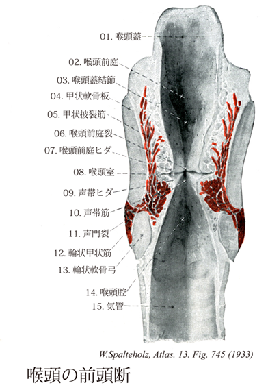
- 745_00【Larynx喉頭 Larynx】 The larynx is situated between the pharynx and trachea.
→(喉頭は咽頭と気管の間にある気道の一部をなすと同時に発声器として重要な役目を有する。前頚部の正中部で、第4~6頚椎の高さにあり、前と外側は皮膚と舌骨下筋群におおわれ、後は咽頭の喉頭部に接する。上部は上後方に向かって咽頭腔に突出し、喉頭口をもって咽頭喉頭部につづく。喉頭は軟骨性の支柱(喉頭軟骨)を有し、声帯ひだを含む弾性膜、およびこれらの位置と緊張を調整する喉頭筋からなる。)
- 745_01【Epiglottis喉頭蓋 Epiglottis】 Elastic cartilage shaped like a shoehorn.
→(喉頭口の前壁をなし、粘膜におおわれて舌根のところにあり、その概形は喉頭蓋軟骨によってつくられ靴べら様を呈する。喉頭蓋の主な働きは燕下を円滑に行うことである。)
- 745_02【Laryngeal vestibule喉頭前庭 Vestibulum laryngis】 It extends from the laryngeal inlet to the vestibular fold.
→(前庭ヒダまでの喉頭腔上部。 (Feneis))
- 745_03【Epiglottic tubercle喉頭蓋結節 Tuberculum epiglotticum】 Prominence in the wall of the epiglottis above the stalk.
→(喉頭蓋茎の上方で、後面の粘膜にある小さな高まり。 (Feneis))
- 745_04【Right/left lamina of thyroid cartilage; Lamina of thyroid cartilage; Thyroid cartilage lamina右板・左板(甲状軟骨の);甲状軟骨板 Lamina cartilaginis thyroideae dextra/sinistra; Lamina dextra/sinistra cartilaginis thyroideae】 Lateral plates of the thyroid cartilage that meet in the midline like the prow of a ship.
→(船首様に正中線で接合されている甲状軟骨の各側板。 (Feneis))
- 745_05【Thyro-arytenoid muscle甲状披裂筋 Musculus thyroarytenoideus; Musculus thyreoarytaenoideus externus】 o: Anterior inner surface of thyroid cartilage, i: Anterior lateral surface of arytenoid cartilage. Synergist in closing the rima glottidis. I: Recurrent laryngeal nerve.
→(甲状披裂筋は甲状軟骨の正中部後面から起こって後走し、披裂軟骨の前外側面に付く。筋線維の一部は声帯靱帯から起こり、靱帯に沿って声帯ヒダのなかを走って披裂軟骨の声帯突起に付き、声帯筋(臨床的に内筋internus)といわれる。神経支配は反回神経。作用は披裂軟骨を前方にひき、声帯ヒダの緊張を減ずる(輪状甲状筋の拮抗作用)。また披裂軟骨の声帯突起内方に回転するので、声門裂を閉じる働きもある。)
- 745_06【Rima vestibuli; Ventricular recess喉頭前庭裂;前庭裂;前庭室裂 Rima vestibuli】 Space between the two vestibular folds.
→(両側の前庭ヒダの間の裂隙。 (Feneis))
- 745_07【Vestibular fold; False vocal cord喉頭前庭ヒダ;前庭ヒダ;室ヒダ;仮声帯 Plica vestibularis; Plica ventricularis】 Fold produced by the vestibular ligament. It lies between the laryngeal ventricle and the laryngeal vestibule.
→(室靱帯によりできたヒダ。喉頭室と喉頭前庭の間に位置する。 (Feneis))
- 745_08Morgagni, Ventricle of【Laryngeal ventricle喉頭室 Ventriculus laryngis】 Lateral outpouching between the vocal folds and vestibular fold.
→(声帯ヒダと前庭ヒダとの間に位置する陥凹。モルガニ洞とも呼ばれる。他にも多数の業績を残すイタリアの医学者Giovanni Battista Morgagni (1682-1771)によって報告された。)
- 745_09【Vocal fold声帯ヒダ Plica vocalis】 The vocal fold overlies the vocal ligament and is supported laterally by the vocalis muscle.
→(喉頭腔のほぼ中央の高さにある2条のヒダの下方にあるもの。声帯ヒダは肉眼的に白く見える。左右の声帯ヒダは前端では合しているが、後端は左右両側の披裂軟骨につくので離れている。基底に声帯靱帯があり、声帯筋により外側から支持されている。声帯ヒダは発声に関する器官。)
- 745_10【Vocalis muscle; Vocal muscle声帯筋;甲状披裂筋の声帯部 Musculus vocalis; Pars vocalis musculus thyreoarytaenoideus】 o: Inner surface of thyroid cartilage near the midline. i: Vocal process and oblong fovea of arytenoid cartilage. Its tensing action changes the intrinsic vibration of the vocal ligament. I: Recurrent laryngeal nerve.
→(甲状披裂筋の筋線維の一部は声帯靱帯から起こり、靱帯に沿って声帯ヒダの中を走って披裂軟骨の声帯突起につく。声帯筋は声帯靱帯から起こるので、声帯ヒダの前部を緊張させ厚みを増す。この作用によって発声時に声帯ヒダの振動部分の長さ・太さを微妙に調節する。)
- 745_11【Rima glottidis声門裂;声帯裂 Rima glottidis; Rima vacalis】 Fissure between the two arytenoid cartilages and the vocal folds.
→(声門裂は声門ヒダによって境された前3分の2の膜間部と、披裂軟骨によって境された後ろ3分の1の軟骨骨部とから構成されている。)
- 745_12【Cricothyroid muscle輪状甲状筋 Musculus cricothyroideus】 o: Anterior and lateral from the cricoid cartilage, i: Inferior margin of the lamina of thyroid cartilage on the inner and outer surface of the inferior horn. If the thyroid cartilage is fixed, it tilts the cricoid cartilage with the arytenoid cartilages posteriorly, thereby tensing the vocal ligaments. I: External branch of superior laryngeal nerve (only one). It is composed in 50% of the following parts.
→(輪状甲状筋は前方で輪状軟骨の弓から起こり、傾斜した内側線維束および、より水平な外側線維束となって甲状軟骨下縁とその下角前縁へ停止する。臨床的に前筋anticusといわれる。作用は甲状軟骨を前方に引き下げる。この筋の作用によって甲状軟骨が前方に傾くと、甲状軟骨正中部の後面と披裂軟骨の声帯突起との間に張る声帯靱帯はややひっぱれて緊張する。すわなち、声帯ヒダの緊張筋である。)
- 745_13【Arch of cricoid cartilage輪状軟骨弓 Arcus cartilaginis cricoideae】 Anterior and lateral part of the cricoid cartilage.
→(輪状軟骨の前部および外側部。 (Feneis))
- 745_14【Laryngeal cavity喉頭腔 Cavitas laryngis; Cavum laryngis】 Space within the larynx.
→(喉頭腔の内面は粘膜におおわれ、下に存在する軟骨、靱帯、筋の形態や走向に従ってヒダや陥凹をつくる。喉頭腔の咽頭喉頭部への開口を喉頭口とよぶ。喉頭口の前部は喉頭蓋、後部は披裂喉頭蓋ヒダに囲まれて楕円形を呈する。喉頭口の後部の左右の披裂軟骨の間で粘膜は観入して披裂間切痕をなす。喉頭口と前庭ヒダの間を喉頭前庭といい、前庭ヒダと声帯ヒダとの間を喉頭室という。喉頭室の盲端は甲状軟骨内面まで伸び、喉頭小嚢となる。サルではこれがよく発達し、響嚢となる。前庭ヒダの支柱は室靱帯で、左右の前庭ヒダとの間の裂隙を前庭裂という。声帯ヒダの下で弾性円錐および輪状軟骨に囲まれた部位を声門下腔という。)
- 745_15【Trachea気管 Trachea】 Elastic tube between the larynx and bronchi.
→(喉頭の下に連なる気道の管状部で、第6頚椎の高さにはじまり、気道の前を垂直に下り、第4頚椎の前で左右の気管支に分岐する。この分岐部を気管分岐部という。気管支鏡で分岐部を上から見ると、その正中部に左右の気管支を隔てる高まりがある。この高まりを気管竜骨という。気管壁には、硝子軟骨性の気管軟骨の輪が一定の間隔をおいて重なり、軟骨間は輪状靱帯で結合する。気管軟骨は幅3~4mmで15~20個を数える。気管軟骨は完全な輪ではなく、全周の4/5~2/3を占める馬蹄状を呈する。軟骨性の支柱を欠く部は正中部後壁をなし、膜性壁とよばれる。膜性壁には平滑筋(気管筋)を含む。気管内面は多列絨毛円柱上皮で、絨毛の運動の方向は上向きである。粘膜固有層には弾性線維が多く、粘膜下組織には胞状の混合腺(気管腺)を数多く含む。日本人の気管の長さは10cm前後である。)