Spalteholz HANDATLAS DER ANATOMIE DES MENSCHEN VON WERNER SPALTEHOLZ
メニューは解剖学(TA)にリンクしてあります。図の番号をクリックすると下記の説明へ、右側の用語をクリックすると解剖学(TA)にジャンプします。
081


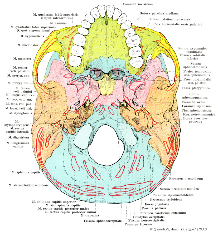
- 081_00【External surface of cranial base外頭蓋底;外面(頭蓋底の) Basis cranii externa】
→(外頭蓋底は下顎骨・舌骨を除く頭蓋の下面をいう。その範囲は上顎骨の歯槽弓にはじまり、側頭下稜、頬骨弓の後端、乳様突起の内側、上項線、外後頭隆起に至る境界とする。外頭蓋底は全体とふくれ上がって凹凸に富み不整複雑な面である。)
- 081_01【Quadratus labii superioris muscle上唇方形筋 Musculus quadratus labii superioris】
→()
- 081_02【Levator labii superioris muscle上唇挙筋;眼窩下筋;眼窩下頭(上唇方形筋の) Musculus levator labii superioris; Musculus infraorbitalis; Caput infraorbitale (Musculus quadratus labii superioris)】 Muscle that arises above the infraorbital foramen and whose fibers insert into the orbicularis oris. I: Facial nerve.
→(上唇挙筋は眼窩下孔の上方で眼窩下縁から上唇に向かって内側下方へ走る。参考:“alaeque nasi”は“et alae nasi”と大体同じ意味。この2筋も、一般に単一筋板を作り、分離しがたい。さらに、小頬骨筋も合わせて3筋を上唇方形筋M. quadratus labii superiorisということがある。)
- 081_03【Levator anguli oris muscle口角挙筋;犬歯筋 Musculus levator anguli oris; Musculus caninus】 Muscle passing from the canine fossa to the angle of the mouth. I: Facial nerve.
→(口角挙筋は犬歯上方および眼窩下孔で犬歯窩から口角まで走る。)
- 081_04【Zygomaticus minor muscle; Zygomatic minor muscle小頬骨筋;頬骨頭(上唇方形筋の) Musculus zygomaticus minor; Caput zygomaticum (Musculus quadratus labii superioris)】 Muscle passing from the zygomatic bone to the upper lip. I: Facial nerve.
→(小頬骨筋は大胸骨筋の内側で起始して上唇に停止する。小頬骨筋は眼輪筋に密着する。小頬骨筋は、上唇挙筋、上唇鼻翼挙筋と協同して、鼻唇溝を深くする(“すすり泣き”)。)
- 081_05【Zygomaticus major muscle; Greater zygomatic muscle大頬骨筋;頬骨筋 Musculus zygomaticus major; Musculus zygomaticus】 Muscle extending from the zygomatic bone to the angle of the mouth and upper lip. I: Facial nerve.
→(大胸骨筋は頬骨弓から内側下方へ向かって上唇および口角まで走る。大頬骨筋は口角を引き挙げ、かつ口角を外方へ引く(狭義の“笑う筋”)。)
- 081_06【Buccinator muscle頬筋 Musculus buccinator】 Muscle arising from the pterygomandibular raphe and adjacent areas of the maxilla and mandible to the height of the first molar teeth, and inserting into the orbicularis oris at the angle of the mouth. It forms the cheek, moves food from the oral vestibule between the dental arcades during mastication, prevents entrapment of the mucous membrane of the mouth, and is active during laughing and crying. I: Facial nerve.
→(頬筋は頬の筋性土台に該当し、口角部で口輪筋に付着する。頬筋は弓状に上顎骨歯槽突起の臼歯部、かつ下顎骨歯槽突起から起こる。上および下顎間は腱性の翼突下顎放線によって橋渡しされ、この放線もまた頬筋の起始である。上咽頭収縮筋の一部がこの放線の後部で起始する。口角付近で、線維索が交叉するので、頬の上方に位置する部分は下唇に広範囲わたって達することもあるし、達しないこともある頬筋は上顎の第2大臼歯のレベルで耳下腺管によって貫通され、しかも本筋は脂肪体からこれを隔てる浅筋膜(頬咽頭筋膜)を有する唯一の顔面筋である。頬筋は上・下歯列弓および頬粘膜間に入り込んだ植物片を再度歯列弓間に押し戻し、咀嚼および植物片のかたちづくりに重要な役割を果たしている。本筋は口腔前庭を圧縮して、空気あるいは液体を口裂を通してふき出す(泡をふき出す、口笛をふく、吐き出す:“トランペット吹きの筋”)。両側の頬筋の収縮はは口角の外側部をくぼませる。参考:この筋は頬粘膜に密に結合しているが、皮膚との間は脂肪組織で隔てられている。上顎第2大臼歯の高さで耳下腺管に貫かれる。)
- 081_07【Masseter muscle咬筋 Musculus masseter】 The most prominent masticatory muscle. It acts to close the mouth and, together with the temporal and medial pterygoid muscles, determines the level of masticatory force. It consists of the following two parts.
→(咬筋は最も浅層にある咀嚼筋である。浅部と深部の2部からなり、浅部は強い腱で頬骨弓の前3分の2の下縁と内面から起こり後下方に向かい、深部は頬骨弓の後3分の2の下縁に垂直に下り向かい下顎枝および下顎角の外面に付く。作用は下顎骨を引き上げて歯をかみ合わせる。咬筋は強大な筋で、歯をかみ合わせると、体表からみることができ、かつ触れることができる。)
- 081_08【Tensor veli palatini muscle口蓋帆張筋 Musculus tensor veli palatini】 o: Spine of sphenoid bone, scaphoid fossa, and anterior (lateral) lip of cartilaginous part of auditory tube, i: After changing direction at the pterygoid hamulus, its fibers merge with the palatine aponeurosis, stiffening the anterior (lateral) wall of the membranous lamina of auditory tube and tensing the soft palate. I: Mandibular nerve.
→(口蓋帆張筋は舟状窩、蝶形骨大翼下面の細い帯および耳管の膜性外壁から起始する。翼突窩のレベルで口蓋帆張筋はすでに腱に移行し、腱は翼突鈎をめぐって方向を転じて、水平に口蓋腱膜へ放射する。口蓋帆張筋は燕下swallowing or deglutionの時に耳管を開く作用がある。)
- 081_09【Lateral pterygoid muscle外側翼突筋 Musculus pterygoideus lateralis; Musculus pterygoideus externus】 o: Lateral surface of lateral plate of pterygoid process and inferior surface of greater wing of sphenoid, i: Two-headed (variant: three-headed) at disco-capsular system of temporomandibular joint and pterygoid fovea. I: Mandibular nerve.
→(外側翼突筋は2頭からなる。上頭は蝶形骨大翼の下面から起こる。下頭は蝶形骨翼状突起外側板に起始する。下頭は側頭下窩を通過して、下顎骨関節突起(翼突筋窩に)停止し、上頭もまた関節円板および関節包に付着する。三叉神経の下顎神経の外側翼突筋神経より支配を受ける。作用として下顎骨を引く。片側が働けば下顎骨前部は対側に働く。)
- 081_10【Medial pterygoid muscle内側翼突筋 Musculus pterygoideus medialis; Musculus pterygoideus internus】 o: Pterygoid fossa and the maxillary tuberosity. i: Pterygoid tuberosity on inner side of the angle of the mandible, passing obliquely downward and backward. Synergist of the temporal and masseter muscles. I: Mandibular nerve.
→(内側翼突筋は蝶形骨の翼突窩で起始して、下顎角内面に停止する。したがって、この筋は、下顎骨の外面側を走る咬筋浅部と同様な走行方向で下顎骨の内側面を走る。両筋は作用方向は同一であり、したがって協力筋である。)
- 081_11【Longus capitis muscle頭長筋 Musculus longus capitis】 o: Anterior tubercles of C3-C6. i: Basilar part of occipital bone. Anterior and lateral flexion of the head and cervical vertebral column. I: Cervical plexus (C1-C3).
→(頭長筋と頚長筋はおうおうにして互いの境界が明瞭に定められない。この筋は縦あるいは斜めに走る線維束を持つ複合羽状構造をもつ。筋腹には通常不完全ながら腱画が挿入されている。頭長筋の起始は第3,4,5,6頚椎横突起の前結節。停止は後頭骨底部の下面。機能として頚椎と糖を屈曲しかつ回旋の補助をする。神経支配は第1,2,3,4頚神経の筋枝。動脈は下甲状腺動脈の上行頚枝、上行咽頭動脈の前脛骨枝、脛骨動脈の筋枝から受ける。)
- 081_12【Rectus capitis anterior muscle; Anterior rectus capitis muscle前頭直筋 Musculus rectus capitis anterior】 o: Lateral mass of atlas, i: Basilar part of occipital bone. Forward bending of the head. I: Cervical plexus (C1).
→(前頭直筋はもっとも頭方に位置し、環椎と後頭骨の間にある。一般に単一分節筋でありC1頚神経前枝のみに支配される。)
- 081_13【Levator veli palatini muscle口蓋帆挙筋 Musculus levator veli palatini】 o: Petrous part of temporal bone in front of the inferior opening of the carotid canal. Inferior border of the cartilaginous auditory tube, i: Palatine aponeurosis. It draws the soft palate backward and upward, also moving the dorsomedial cartilaginous part of auditory tube when the pharyngeal opening of auditory tube is opened. I: Vagus nerve.
→(口蓋帆挙筋は、口蓋帆張筋の後に位置し、側頭骨錐体部下面で頚動脈管の前部および耳管軟骨から起始する。口蓋帆挙筋は耳管に沿って斜め前・下方へ走り、軟口蓋にはいる。両側の口蓋帆挙筋の腱線維はからみ合って、高さが調節できる筋性ワナを形成する。)
- 081_14【Styloglossus muscle茎突舌筋 Musculus styloglossus】 o: Styloid process, i: Radiates from posterosuperior into the lateral part of the tongue and merges with the hyoglossus. It draws the tongue backward and upward. I: Hypoglossal nerve.
→(茎突舌筋は外舌筋の1つ。茎状突起(および茎突下顎靱帯)から放射して口蓋咽頭弓のレベルで舌に至る。茎突舌筋の線維の主部は舌縁で舌尖に向かって走り(筋の縦索)、個々の線維束は内側へ曲がり、横舌筋(内舌筋)の線維に付着する。)
- 081_15【Stylopharyngeus muscle茎突咽頭筋 Musculus stylopharyngeus】 o: Styioid process, i: It extends medially between the superior and middle constrictor muscles and reaches the wall of the pharynx, thyroid cartilage, and epiglottis. I: Glossopharyngeal nerve.
→(茎突咽頭筋は茎状突起の頭蓋底近くから起こり、上および中咽頭収縮筋の間を通って筋の内面に至り、口蓋咽頭筋の線維束とともに甲状軟骨に停止する。一部は咽頭蓋の粘膜下に終わる。)
- 081_16【Rectus capitis lateralis muscle; Lateral rectus capitis muscle外側頭直筋 Musculus rectus capitis lateralis】 o: Transverse process of atlas, i: Jugular process of occipital bone. Lateral bending of the head. I: Anterior ramus of spinal nerve (C1).
→(外側頭直筋は椎間横突起の前部に起こり、後頭骨頚静脈突起に停止する。C1頚神経前枝に神経支配される。作用として頭を同側に曲げる。)
- 081_17【Digastricus muscle; *Digastric muscle顎二腹筋 Musculus digastricus; Musculus biventer mandibulae】 o:Mastoid notch, i: Digastric fossa. It has an intermediate tendon that acts on the lesser horn of the hyoid bone by means of a connective tissue sling. Raises the hyoid bone and opens the mouth.
→(顎二腹筋は舌骨の上方にある細長い筋で中間腱で前腹と後腹との2腹に分かれる。その後腹をもって側頭骨乳突切痕で起始し、斜め前・下方へ走る。舌骨付近で後腹は中間腱に移行し、この腱は二分した茎突舌骨筋によって挟まれ、かつ線維性滑車によって舌骨に固定される。前腹(顎舌骨筋からは皮膚側へ位置しているが)は中間腱から起始し、下顎骨内面で下顎下縁近くの二腹筋窩に停止する。顎二腹筋の前腹(下顎神経の枝である顎舌骨筋神経の支配)と後腹(顔面神経の支配)とは神経支配が異なることは注意を要する。下顎が固定されているときには、舌骨を引き上げる。舌骨が固定されているときは下顎骨を後下方に引く。両者は発生学的にも由来を異にし、前腹は顎舌骨筋・口蓋帆長筋などとともに咀嚼筋と同類(鰓弓のうち顎骨弓mandibular archに属する筋)であり、後腹は茎突舌骨筋・アブミ骨筋などとともに顔面表情筋と同類(鰓弓のうち舌骨弓hyoid archに属する筋)である。ちなみに、咀嚼筋は下顎神経で支配され、顔面表情筋は顔面神経支配である。このように発生学的な由来を知れば、色々な筋の支配を整然と整理することができる。)
- 081_18【Longissimus capitis muscle頭最長筋 Musculus longissimus capitis】 o: Transverse processes from T3-T1 and C7-C3. i: Mastoid process.
→(頭最長筋は「最長筋の頚部起始筋束の内側で」C3~T3の横突起から起始する。この筋線維は細長い、より矢状方向に配列した筋束を形成し、側頭骨の乳様突起に停止する。)
- 081_19【Splenius capitis muscle頭板状筋 Musculus splenius capitis】 Portion of the splenius extending to the head. o:Spinous processes of T3-C4. i: Lateral part of the superior nuchal line and mastoid process.
→(頭板状筋の起始は頚靱帯の下半分、第7頚椎と上位3~4個の胸椎棘突起。停止は側頭骨の乳様突起と上項線の外側部。機能として共同で頭と頚の伸展と側方屈曲をしかつ頭を少し回旋する。神経支配は中および下頚神経の後枝の外側枝。動脈は後頭動脈下行枝の筋枝、頚横動脈の浅枝から受ける。)
- 081_20【Sternocleidomastoid muscle胸鎖乳突筋 Musculus sternocleidomastoideus】 o: Two-headed muscle arising from the sternum and clavicle, i: Mastoid process; superior nuchal line. Rotates the face to the contralateral side and bends the head to the ipsilateral side. Bilateral contraction elevates the face. I: Accessory nerve, cervical plexus (C1-C2).
→(胸鎖乳突筋は側頚部にある強大な斜めに縦走する浅層の筋。胸骨柄前面と鎖骨の胸骨端から2頭をもっておこり、両頭は合して強い筋腹をつくって後上方に走り、乳様突起および後頭骨の上項線につく。作用は複雑で、両側が同時に働くとオトガイを上げて後頭部を片側が働けば頭を対側にまわすが、その浅オトガイが対側に向かって上り、頭は逆に同側に傾く。支配神経は副神経外枝と頚神経叢筋枝(C2, C3)であり、したがって僧帽筋と同系の筋である。また、第6咽頭弓に発生する鰓弓筋で、鎖骨上窩を囲む2頭(胸骨頭と鎖骨頭)をもって始まる。胸骨頭は胸骨柄の上縁から、鎖骨頭は鎖骨の胸骨端から起こる。筋膜は頚筋膜浅葉に鞘状に包まれており、斜め上方に向かって幾分螺旋状に回転しながら頚部外側面を横切り、よく発達した腱となって乳様突起と上項線に停止する。筋の表面は、起始部で腹側に、停止部で外側に向く。参考:副神経外枝の僧帽筋枝は、外枝がこの筋に入る前に分かれることと、筋内で分かれて再び外に現れることがある。胸鎖乳突筋はドイツ語ではKopfnicker(頭をこっくりとうなずかせる筋)と呼ばれるが、これは作用の点からは正しくない。この筋が片側だけ収縮すると、頭はその側へ傾き反対側を振り向いて、あたかも「首をかしげる」状態になる。また両側の物が同時に収縮すると、頭を胴体にめり込ませるように働くのえある。Musculus sternocleidomastoideusというラテン名はあまりにも長たらしいので、米英では多少簡略化してsternomastoid muscleともよぶ。片側の胸鎖乳突筋が先天的に短い場合、または出産時の外傷などによって瘢痕化して短縮すると、この筋の作用を考えればすぐわかるように、頭は病側へ傾くと共に健側にねじれたままの状態になるこれを斜径torticolis, wryneck(性格には筋性斜径)といい、かなり頻度の高いものである。略語(SCM))
- 081_21【Obliquus capitis superior muscle上頭斜筋;頭斜筋 Musculus obliquus capitis superior; Musculus obliquus capitis】 o:Transverse process of atlas, i: Area overlying the insertion of the rectus capitis posterior major. Posterior and lateral bending of the head. I: Suboccipital nerve.
→(上頭斜筋は、もっとも上方に位置する後横突間筋であり、環椎の横突起の後部から下項線まで少々上向きに走行し、大後頭直筋の停止部の少々上で、外側部に停止する。)
- 081_22【Semispinalis capitis muscle頭半棘筋;横突後頭筋 Musculus semispinalis capitis; Musculus transversooccipitalis】 o: Transverse processes of T6-C3. i: Inferior to the superior nuchal line. I: Posterior rami of spinal nerves of C1-C5.
→(頭半棘筋の起始は上位6個の胸椎と第7頚椎の横突起、第4~6頚椎関節突起。停止は後頭骨の上下項線間の項平面。機能として脊柱の伸展、側方屈曲。頭、肋骨、骨盤の伸展。神経支配は第1~6頚椎。動脈は後肋間動脈の筋枝、後頭動脈の下行枝、肋頚動脈の深頚枝から受ける。頭半棘筋は頚部の板状筋に完全におおわれ頚最長筋と頭最長筋の内側にある。固有背筋の外側筋群を形成する筋原基から大部分形成される。それ故に、この筋は脊髄神経後枝の内側枝ばかりでなく、外側枝の支配も受ける。この筋は複合羽状型であり、不完全に狭い内側筋束と、線維質の外側筋束に分化し、両者とも中間腱を所有する(内側筋束はときどき2つ)。)
- 081_23【Rectus capitis posterior major muscle大後頭直筋 Musculus rectus capitis posterior major】 o:Spinous process of axis, i: Middle of inferior nuchal line. Rotates the face laterally. Posterior flexion. I: Suboccipital nerve.
→(大後頭直筋は軸椎の棘突起から外側方向に下項線の中1/3の上方に走行する。作用:主として頭を後ろに引いて直立位に保持する。一側が動けば同側にまげる。また下頭斜筋は同側に回す。参考:後頭下筋は回旋筋などの最上部に相当するが、上頭斜筋だけは神経支配を異にし、最長筋系に属する。)
- 081_24【Rectus capitis posterior minor muscle; Minor posterior rectus capitis muscle小後頭直筋 Musculus rectus capitis posterior minor】 o:Posterior tubercle of the posterior arch of atlas, i: Medial one-third of inferior nuchal line. Mainly posterior flexion of the head. I: Suboccipital nerve.
→(小後頭直筋の短筋(もっとも上方に位置する後ろ横突間筋)は環椎後隆起で起始し、下項線の下で大後頭直筋の内側に停止する。)
- 081_25【Trapezius muscle僧帽筋 Musculus trapezius】 Muscle that consists of three parts that act together to position the scapula and clavicle, draw both toward the vertebral column, and brace the shoulder girdle. I: Accessory nerve; brachial plexus C2-C4.
→(背部第1層にみられる扁平な菱形の筋で背部上半部を占める。僧帽筋は上肢の運動の時に肩甲骨を動かす重要な筋である。とくに上腕の外転のときに、肩甲骨を後内側に引くと同時に下角を外側に回し、関節窩が上外側を向くようにする。僧帽筋は下行部、横走部、上行部に分けられる。[臨床]僧帽筋の完全麻痺(副神経と上部腕神経の同時の傷害)の場合、肩は健側よりも深く位置するようになる項肩線は弓状を呈さず、乱れる。肩甲骨は正中線より、はるかに離され、関節窩は前下方を向く。肩は(肩甲挙筋の)弱いエネルギーにより持ち上げることが出来るにすぎず、わずかに(菱形筋により)後方にもたらされるにすぎない。腕の外側への挙上は大きく減少する。腕は通常水平面まで外転され得ない。腕の前方への挙上は(前鋸筋による肩甲骨の回転により)ほとんど制限さされないが、矢状面での挙上は強く妨げられる。副神経のみが傷害された場合、僧帽筋の下行部の機能は(上頚神経の付随的支配により)種々の程度に保存される。肩甲骨の位置の変化はそれほど著明ではない。しかし、腕を横または後へ挙上することは、ちょうどその程度に応じて制限される。)
- 081_26【Sphenooccipital fissure蝶後頭裂 Fissura sphenooccipitalis】
→(")
- 081_27Stenson's foramen【Incisive foramina切歯孔 Foramen incisiva】 Between two and four openings of the incisive canals.
→(切歯窩の中に両側の切歯管が切歯孔をもって開く。その上端は鼻稜の前方部の両側で鼻腔に開く。鼻稜のこれより前、すなわち切歯骨の領域にある部は切歯稜ともよばれる。)
- 081_28【Median palatine suture正中口蓋縫合 Sutura palatina mediana】 Suture visible from the oral cavity joining the two halves of the palatine bone.
→(正中口蓋縫合は骨口蓋の左右半部の間の縫合で、鼻腔面に鼻稜をつくる。)
- 081_29【Transverse palatine suture横口蓋縫合 Sutura palatina transversa】 Suture between the palatine process of the maxilla and the palatine bone.
→(横口蓋縫合は骨口蓋の後部を横走し、上顎骨がつくる部と口蓋骨がつくる部との間の縫合。)
- 081_30【Horizontal plate of palatine bone水平板;口蓋板;水平部(口蓋骨の) Lamina horizontalis; Lamina palatina; Pars horizontalis (Os palatinum)】 It forms the posterior third of the hard palate and thus the floor of the nasal cavity.
→(水平板は上顎骨口蓋突起をうしろに延長して骨口蓋をつくる上部で、上面(鼻腔面)は滑らかで、他側のものと会する縁は上顎骨におけると同じく高まり(鼻稜)、さらにうしろに向かって突出する(後鼻棘)。下面(口蓋面)は粗面で、へこみ、前縁にときに高まり(口蓋稜)がみられ、外側縁後方に大口蓋孔がある。)
- 081_31【Zygomaticomaxillary suture頬骨上顎骨縫合 Sutura zygomaticomaxillaris】
→(眼窩下縁の上壁で同管内方に境界にあたり、細い眼窩下縫合があって、眼窩下孔までつづく。これは上顎体と頬骨突起との癒合したあとであるが、老人では明らかでない。)
- 081_32【Inferior orbital fissure下眼窩裂 Fissura orbitalis inferior】 Opening between the greater wing of the sphenoid and the orbital surface of the maxilla. It transmits the zygomatic nerve, infraorbital nerve, and accompanying vessels.
→(眼窩の下壁と外側壁との間に長く大きな下眼窩裂(蝶形骨大翼と上顎骨眼窩部の間の裂隙)がある。下眼窩裂は側頭下窩・翼口蓋窩に通じ、眼窩下動・静脈、眼窩下神経、頬骨神経などの通路となる。眼窩裂の前内側縁から前方に向かって深い溝が出る。この溝を眼窩下溝といい、さらに下壁の下を走って眼窩下縁の下方で眼窩下孔となって上顎骨の前面に開く。)
- 081_33【Sphenofrontal suture蝶前頭縫合;蝶骨前頭縫合 Sutura sphenofrontalis】 Suture that gradually ascends posteriorly along the lateral aspect of the cranium, joining the greater wing of the sphenoid and the frontal bone. Interior cranium: suture that joins the frontal bone and the lesser wing of the sphenoid.
→(蝶前頭縫合は頭蓋の外側面で、蝶形骨大翼と前頭骨の間を孔へゆるやかに登る縫合線。頭蓋内面では、前頭骨と蝶形骨小翼の間にある。)
- 081_34【Temporal surface of sphenoid側頭面(蝶形骨の) Facies temporalis (Ossis sphenoidalis)】 Lateral facing surface of the greater wing of the sphenoid.
→(蝶形骨大翼の外方を向く面は側頭面で、その大部分は頭蓋の側面において側頭窩の底となる。)
- 081_35【Pyramidal process of palatine bone錐体突起(口蓋骨の) Processus pyramidalis (Os palatinum)】 The inferoposterior end of the perpendicular plate of the palatine bone, which is inserted in the pterygoid notch.
→(垂直板の下部は水平板より矢状径が広くなり、水平板より後に大きく突出する錐体突起となって蝶形骨翼状突起の翼突切痕にはまる。)
- 081_36【Pterygoid fossa翼突窩 Fossa pterygoidea】 Depression between the lateral and medial plates of the pterygoid process for the medial pterygoid muscle.
→(翼状突起の内側板、外側板は後方に開いた翼突窩をつくる。ここから内側翼突筋が起始する。)
- 081_37【Sphenosquamosal suture蝶鱗縫合;蝶骨鱗縫合 Sutura sphenosquamosa】 Suture between the squamous part of the temporal bone and the greater wing of the sphenoid.
→(蝶鱗縫合は側頭骨鱗部と蝶形骨大翼の間の縫合。)
- 081_38【Foramen ovale of sphenoid bone卵円孔(蝶形骨の) Foramen ovale】 Opening for the passage of the mandibular nerve anteromedial to the foramen spinosum.
→(卵円孔は大翼の後内側端に位置し、三叉神経の下顎神経の通路の開口で、棘孔の内前方にある。海綿静脈洞と翼突静脈叢を連絡することがある。)
- 081_39【Foramen spinosum棘孔(蝶形骨の) Foramen spinosum】 Opening posterolateral to the foramen ovale for the passage of the middle meningeal artery.
→(中硬膜動脈の通路の開口で、卵円孔の外後方にある。(Feneis))
- 081_40【Petrosphenoidal fissura; Sphenopetrosal fissure蝶錐体裂;蝶骨錐体裂 Fissura sphenopetrosa】 Prolongation of the petrosquamous fissure to medial that is continuous with the foramen Iacerum. Passage of the lesser petrosal nerve and exit of the chorda tympani from the cranium.
→(蝶錐体裂は破裂孔から後外方に、大翼と錐体の間にある。蝶錐体裂の内後方にある大錐体神経溝の前端は破裂孔に達し、小錐体神経溝は小錐体神経溝は蝶錐体裂の後端から卵円孔に向かう。)
- 081_41Glaserian fissure グラーザー裂【Petrotympanic fissure錐体鼓室裂 Fissura petrotympanica】 (glaserian fissure). Fissure located posteromedial to the mandibular fossa between the tympanic part and the visible strip of the petrous part of the temporal bone. Its medial part can lodge the chorda tympani.
→(鼓室部の前上縁は下顎窩(顎関節の関節窩)の後縁にある錐体鼓室裂である。)
- 081_42【External acoustic opening外耳孔;骨外耳孔 Porus acusticus externus】 Opening to the external acoustic meatus.
→(外耳道を作るときに前後両縁が中央部より速く発育し、その尖端で癒合するために、ある時期には外耳道下壁の骨板に孔を有することがある。)
- 081_43【Mastoid foramen乳突孔 Foramen mastoideum】 Opening behind the mastoid process for transmission of the mastoid emissary vein.
→(乳突様突起の後方にある孔。小動脈を硬膜へ、導出静脈をS状静脈洞へ送っている。)
- 081_44【Occipitomastoid suture後頭乳突縫合 Sutura occipitomastoidea】 Continuation of the lambdoid suture to the cranial base.
→(後頭乳突縫合は後頭骨と側頭骨乳突部の間。)
- 081_45【Stylomastoid foramen茎乳突孔 Foramen stylomastoideum】 External opening of the facial canal behind the styloid process and between the mastoid process and the jugular fossa.
→(錐体下面の後外側端は茎状突起の着く所で、これとそ後方の乳様突起との間にある茎乳突孔は顔面神経管の出口である。)
- 081_46【Styloid process of temporal bone茎状突起(側頭骨の) Processus styloideus (Ossis temporale)】 Long bony process in front of the stylomastoid foramen. It is a relic of the hyoid arch.
→(茎状突起は錐体下面の後外側端から前下方へ向かう細長い突起である。その長さは1~5cmで、茎突下顎靱帯、茎突舌骨靱帯、茎突喉頭筋などの起点となる。茎状突起の根部の前面は茎状突起鞘で被われる。なお、茎状突起は舌骨と関係ある第2鰓弓軟骨の一部が骨化したも野である。)
- 081_47【Jugular fossa頚静脈窩;頚窩 Fossa jugularis】 Widening of the jugular foramen that contains the superior bulb of the jugular vein.
→(錐体下面の後縁に近い中部には弓状の大きく深い頚静脈窩がる。頚静脈上球を容れる。)
- 081_48【Petrosal fossula錐体小窩 Fossula petrosa】 Small depression on the ridge between the carotid canal and the jugular fossa for the tympanic ganglion of glossopharyngeal nerve.
→(頚動脈管外口と頚静脈かとの間には三角形の小さい凹みがあるが、これは舌咽神経の下神経節を容れる錐体小窩である。錐体小窩の底には細い鼓室小管が開く。)
- 081_49【External opening of carotid canal頚動脈管外口;外口(頚動脈管の) Apertura externa (Canalis caroticus); Foramen caroticum exernum】 Opening in the external cranial base between the jugular foramen and the musculotubal canal.
→(頚静脈下の前内側には大きい頚静脈管外口がある。この頚静脈管外口の前から口蓋帆張筋の一部が起こる。)
- 081_50【Occipital condyle; *Condyle後頭顆 Condylus occipitalis】 Spherical eminence for articulation with the atlas.
→(後頭骨下面にある2つの細長い卵形をした関節面を有する高まりが後頭顆である。これは第1頚椎の上関節窩と関節する。)
- 081_51【Petro-occipital fissure錐体後頭裂 Fissura petrooccipitalis】 Medial continuation of the jugular foramen that extends from the temporal bone to the occipital bone.
→(錐体後頭裂は岩様部(錐体)と後頭骨の間にあり、頚静脈孔のつづきとして、頚静脈孔の上方に位置する。これに一致して下錐体洞溝がある。)
- 081_52【Foramen lacerum破裂孔 Foramen lacerum】 Irregularly bordered aperture that is closed with fibrocartilage. It is located between the apex of the petrous part of the temporal bone and the sphenoid in the middle cranial fossa and gives passage to the deep petrosal nerve and the greater petrosal nerve.
→(破裂孔は生体では軟骨で閉ざされている。破裂孔はその前縁の一部が蝶形骨で形成され、その部位より内頚静脈は海綿静脈洞に入る。)