Spalteholz HANDATLAS DER ANATOMIE DES MENSCHEN VON WERNER SPALTEHOLZ
メニューは解剖学(TA)にリンクしてあります。図の番号をクリックすると下記の説明へ、右側の用語をクリックすると解剖学(TA)にジャンプします。
761


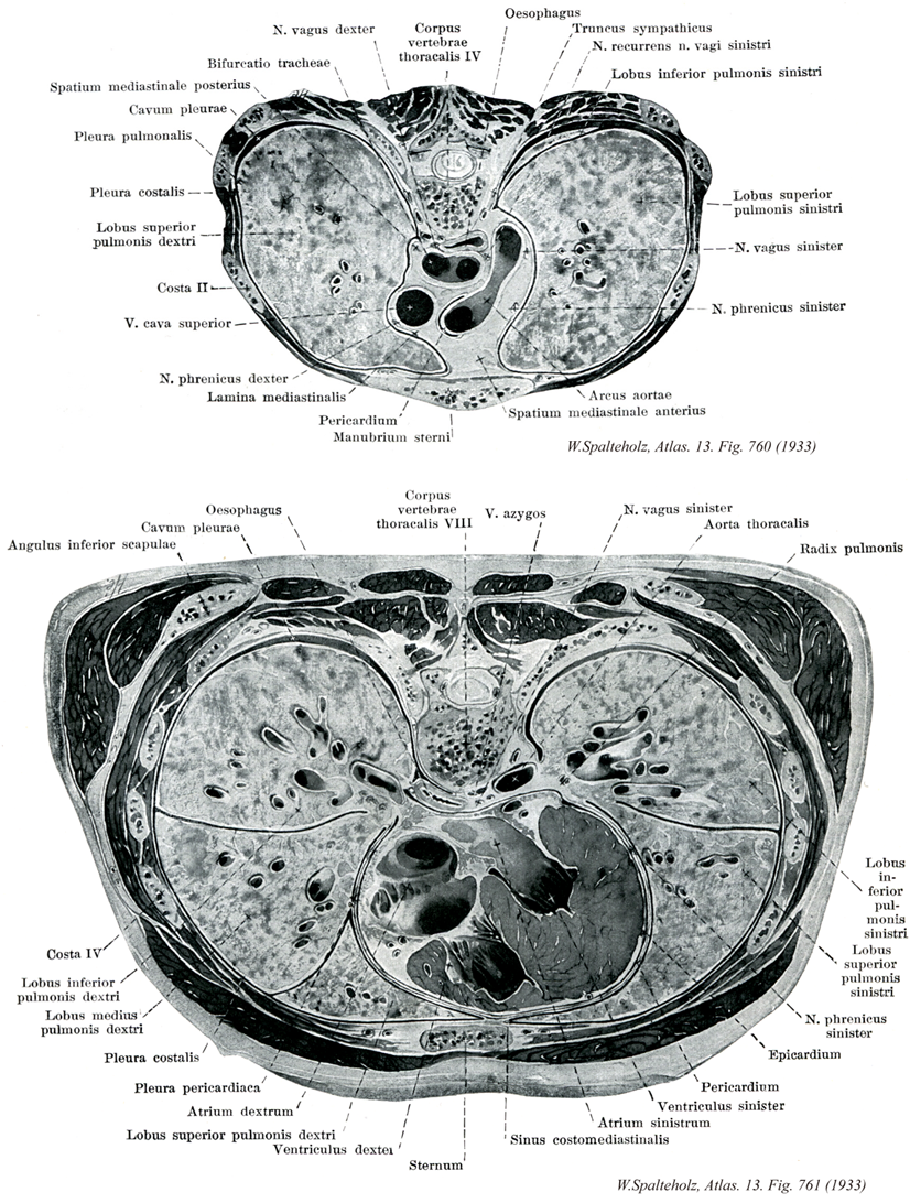
- 761_01【Body of 8th thoracic vertebra; Eighth thoracic vertebral body第8胸椎椎体 Corpus vertebra (Vertebra thoracic VIII)】
→()
- 761_02【Oesophagus; Esophagus食道 Oesophagus; Esophagus】 Passageway measuring 23-26 cm in length that begins below the cricoid cartilage at the level of the sixth cervical vertebra and ends at the cardia of the stomach.
→(食道は咽頭につづき、下方は胃に流入する長い管で、狭義の消化管の最初の部分である。輪状軟骨下縁(上食道狭窄)にはじまり、脊柱の前を下って胃の噴門部に接合するまで、全長23~26cm。内腔は適宜拡がり、義歯を飲み込んだ例もある。内腔の狭い部分は上端(上食道狭窄)、大動脈弓・気管支と交叉する部分(中食道狭窄)、下端(下食道狭窄)の3カ所で、上下端では内腔が普通は閉じ、括約筋の存在が想定されている。食道を上から頚部・胸部・腹部に分ける。頚部は脊椎の前にある部分、胸部は以下横隔膜で、腹部は横隔膜の食道裂孔を抜けて腹腔内に入り、噴門部に流入する短い部分である。食道の壁の粘膜は重層扁平上皮におおわれ、粘膜筋板を有し、食道腺が散在する。上部または下端に食道噴門腺をみる。筋層は上部で横紋筋、下部で平滑筋で、平滑筋束の一部は気管支食道筋、胸膜食道筋として、周囲の器官に連続する。筋層の外側は疎性結合組織性の外膜におおわれる。)
- 761_03【Pleural cavity胸膜腔 Cavitas pleuralis; Cavum pleurae】 Slitlike space between the parietal and visceral pleura containing a capillary layer and a small amount of serous fluid.
→(胸膜腔とは、壁側胸膜とその続きである肺胸膜によって囲まれる閉じた空間であって、その内部に存在するのは少量の液(漿液serous fluid)だけである。肺は胸腔の中にあるけれども、胸膜腔の外にある。胸腔の中には心膜によて囲まれる心膜腔という閉じた袋もあり、これらに介在して胸部内臓や大血管などが胸腔内に詰まっているわけである。)
- 761_04【Inferior angle of scapula下角(肩甲骨の);肩甲骨下角 Angulus inferior scapulae】 Lower angle of the scapula.
→(肩甲骨の三角形の三つの頂角のうち下方のものが下角である。)
- 761_05【Fourth rib [IV]第4肋骨 Costa IV; [IV]】
→()
- 761_06【Inferior lobe; Lower lobe of lung下葉(肺の) Lobus inferior pulmonis】 It mainly extends dorsally. Its superior border runs obliquely from posterosuperior to anteroinferior. It begins paravertebrally at the fourth rib and ends at the intersection of the middavicular line and the sixth rib.
→(後側に主な拡がりをもつ。その上限界上後方から下前方へ斜めに走り、第四肋骨から脊柱側方で鎖骨中間線を第六肋骨が切る線までいたる。 (Feneis))
- 761_07【Middle lobe of right lung中葉[右肺の] Lobus medius (Pulmo dexter)】 Present only in the right lung. It lies in front of the midaxillary line between the fourth and sixth ribs.
→(右側のみにある。第四肋骨と第六肋骨の間、腋窩中間線より前に位置する。 (Feneis))
- 761_08【Costal part of parietal pleura; Costal pleura肋骨胸膜;肋骨部(壁側胸膜の) Pars costalis pleurae parietalis; Pleura costalis】 Part of the parietal pleura that lies on the inner surface of the thoracic wall.
→(壁側胸膜の肋骨胸膜は、胸壁の内面(胸骨、肋軟骨、肋骨、肋間筋、肋間膜、胸椎の側面)を覆う。)
- 761_09【Pericardiac part of pleura心膜部(胸膜の);心嚢胸膜 Pars pericardiaca (Pleura parietalis); Pleura pericardiaca】
→(")
- 761_10【Right atrium右心房 Atrium cordis dextrum; Atrium dextrum】
→(右心房は心臓の右上部を占め、その後上部と後下部とに、それぞれ、上大静脈と下大静脈が注いでいる。)
- 761_11【Right superior lobe of lung上葉(右肺の) Lobus superior (Pulmo dexter)】
→()
- 761_11a【Superior lobe of lung; Upper lobe of lung上葉(肺の) Lobus superior pulmonis】 Superior lobe that extends posteriorly to the fourth rib. In the right lung, its inferior border runs anteriorly at about the level of the fourth rib. In the left, its inferior border extends to the osseocartilaginous border of the sixth rib.
→(後方では第四肋骨まで達する。右側では、その下端は第四肋骨にほぼ沿って、前方へいたる。左側では、第六肋骨の骨軟骨境界まで達する。 (Feneis))
- 761_12【Right ventricle右心室 Ventriculus dexter】
→(右心室は心臓の最下位部を占め、後上方にある右房室口で右心房と交通し、前上方にある肺動脈口で肺静脈に連なる。)
- 761_13【Sternum胸骨 Sternum】
→(胸骨は第一から第七肋骨の肋軟骨と鎖骨の軟骨に結合し、胸前壁の中心部を形成する長く平らい骨。胸郭前壁正中部にある縦長の扁平骨で、胸骨柄、胸骨体、剣状突起からなる。胸骨柄は最も側頭にあり、不整六角形を呈す。上縁正中部の浅い陥凹部が頚切痕で、この外側下方で斜め上方に向かう浅い陥凹部が、鎖骨関節面に対する鎖骨切痕である。鎖骨切痕は下方では左右の幅が尾側ほど狭くなり、下縁で胸骨体と軟骨結合によって連結している。胸骨体は縦長の長方形を呈し、下方でやや幅が広い。内面は比較的平滑であるが、外面は分節的に発生した名残として、横走する隆起線が肋骨切痕に対応して数本認められる。胸骨柄と胸骨体の外側縁の浅い陥凹部が、第1~第7肋軟骨に対する肋骨切痕である。第1肋骨切痕は鎖骨切痕の下方にあり、第2肋骨切痕は胸骨柄と胸骨体との連結部で両方にまたがっている。第3肋骨切痕以下は胸骨体にあるが、第5肋骨切痕以下では下方ほど間隔が狭くなる。第7肋骨切痕は剣状突起上端部に接している。剣状突起は胸骨体下縁に接する細長い小部で、一部が軟骨で、形は不定である。胸骨柄と胸骨体(胸骨柄結合)および胸骨体と剣状突起(胸骨瞼結合)の軟骨結合部は、年齢とともに骨化する。胸骨柄結合部は前方にやや突出し、胸骨角をなす。頚切痕外側部で時に見られる小骨が胸上骨である。ギリシャ語のsternon(男性の胸)に由来する。)
- 761_14【Azygos vein奇静脈;右縦胸静脈 Vena azygos; Vena thoracica longitudinalis dextra】 Vein lying on the vertebral column that begins at the ascending lumbar vein and opens at the level of the fourth to fifth thoracic vertebrae into the superior vena cava before it enters the pericardium.
→(奇静脈は主として胸壁および後腹壁よりの静脈血を集めて脊柱の右側を上行する静脈で、第1または第2腰椎の高さで無束の上行腰静脈からつづいてはじまり、横隔間君お右脚を貫いて胸腔に入り、脊柱の右側に沿って上行したのち、第4胸椎の高さで右気管支の上をこえて前方にまがり、上大静脈にそそぐ。発生学的には胎生早期に体壁・下半身および中腎よりの血液を集めた後主静脈に由来する。のち中腎の退化、主上静脈と後主静脈の近位部から生ずる。奇静脈・半奇静脈・副半奇静脈(上行腰静脈)を総称して奇静脈系azygos ssytemといい、大静脈系caval systemに通過障害が起こった場合の側副路の一つとして重要である。奇静脈系は変異に富み、奇静脈・半奇静脈・副半奇静脈が全部そろって存在するのは35%にすぎず、上記のいずれか一つが欠場する場合(30%)や、奇静脈だけが存在する場合(35%)もある。Azygosというラテン語は、a(否定)zygos(2頭の牛の首にわたす軛(くびき)すなわち対をなすもの)、つまり対をなさないという意味である。奇静脈の奇は、偶数に対する奇数の奇であって、奇妙の奇ではない。)
- 761_15【Vagus nerve [X]迷走神経[脳神経X] Nervus vagus [X]】 Nerve arising from the fourth and fifth pharyngeal arches. It emerges from the medulla oblongata together with CN IX in the posterolateral sulcus and passes through the jugular foramen. Its distribution area extends into the thoracic and abdominal cavities.
→(迷走神経は第10脳神経で、上方の舌咽神経、下方の副神経の間で延髄の外側から多数の小根によって起こる混合神経で胸腹部の諸内臓に分布する副交感神経節前神経線維(延髄迷走神経背側核に細胞体をもつニューロンの神経突起)を主成分としている。これらの線維が胸腹部を走行するあいだに、きわめてしばしば自律神経叢を形成してどこに神経の本幹が存在するか不明瞭となるため、迷走神経の名がつけられた。また迷走神経には胸腹部の内臓の知覚を伝える神経線維(その細胞体は迷走神経の下神経節内に存在する)、咽頭下部および後頭の筋への運動線維(延髄疑核に発し、咽頭に分布するものは舌咽神経からの枝とともに咽頭壁において咽頭神経叢を形成したのち筋に分布する)、咽頭下部および後頭の粘膜への知覚神経線維、などが含まれる。後頭に分布する運動および知覚神経線維は下神経節の直下で後頭に向かう上喉頭神経となるか、あるいは胸腔内で迷走神経本幹から下喉頭神経として分かれて頚部を反回神経として上行するかして目的の器官に達する。)
- 761_16【Thoracic aorta胸大動脈;大動脈胸部 Pars thoracica aortae; Aorta thoracica】 Part of the aorta descending to the aortic hiatus of the diaphragm at the level of the twelfth thoracic vertebra.
→(胸大動脈は、大動脈弓の延長である。第4胸椎体の下縁の左側で始まり、第5から第12胸椎の左側で後縦隔を下行する。下行しながら、正中面に近付き、食道と脊柱の左側に沿って走るが、食道の後方、脊柱の前を走るようになる。胸大動脈は横隔膜を貫いた直後に腹大動脈という名前に変わる。胸大動脈と腹大動脈とを総称して下行大動脈という。)
- 761_17【Root of lung; Lung root肺根 Radix pulmonis】 It is composed of the main bronchus, blood vessels, lymph vessels and nodes, and autonomic plexuses.
→(肺根は肺門部出入りするすべての構造をさし、胸膜に包まれて脚状をなす。気管支、肺動静脈、気管支動静脈、リンパ管、神経を含む。)
- 761_18【Phrenic nerve横隔神経 Nervus phrenicus】 Nerve arising from C4 with accessory branches from C3 and C5. It lies on the anterior scalene muscle and then passes anterior to the hilum of lung to the diaphragm, with some fibers continuing into the peritoneum.
→(第3~5頚神経から出て頚神経叢を形成し、主に第4頚神経から起こる。頚部では前斜角筋の前面に沿って、また胸腔中では縦隔胸膜と心膜との間を通って、それぞれ走行する。横隔膜にいたる運動神経であるが、壁側縦隔胸膜、心膜、横隔胸膜、腹膜に知覚神経を送り(心臓枝)、腹腔神経叢からの枝と交通する(横隔腹枝)。時に鎖骨下筋神経または腕神経叢の他の神経から小枝が出てて、第1肋骨付近の高さで横隔神経に合することがあるが、これを副横隔神経という。)
- 761_19【Visceral layer of serous pericardium; Epicardium臓側板;臓側葉(漿膜性心膜の);心外膜 Lamina visceralis pericardii; Epicardium】 Serous covering of the heart. It transitions into the parietal layer near the great vessels.
→(心外膜は心臓表面の漿膜性被覆。中皮と、線維の豊富な固有層からなる。)
- 761_20【Pericardium心膜 Pericardium】 Lubricant-containing sheath enclosing the heart. It consists of a fibrous layer and a double-layered serous coat.
→(心膜は心臓と大血管起始部の被覆と活動のための膜。外層の線維性心膜fibrous pericardiumと内層の漿膜性心膜serous pericardiumの2層からなる閉鎖嚢。漿膜性心膜は心臓表面を直接おおう臓側板(心外膜)と線維性心膜の内面をおおう壁側板にわけられる。線維性心膜は強靱な膜で、大血管の壁につづき、心臓を固定・保持するとともに、その急激な過度の拡張を防ぐ。さらに心臓は心膜腔で囲まれ、潤滑な心膜性心膜で包まれるので、摩擦なく拍動することができる。)
- 761_21【Left ventricle左心室 Ventriculus sinister】
→(左心室は心臓の左下部を占め、後上方にある左房室口で左心房と交通し、右上隅にある大動脈口によって大動脈につらなる。左心室の壁は右心室に比べ2~3倍厚い。心室中隔は、右心室に向かって膨隆しているので、心室を横断面でみると、左心室の内腔は円いのに対して、右心室の内腔は半月状である)
- 761_22【Left atrium左心房 Atrium cordis sinistrum; Atrium sinistrum】
→(左心房は心臓の後上部にあって、後面をつくっている。左心房は右心房よりもやや小さいが、壁はやや厚い。左心房の後壁の上部に、左右両肺からそれぞれ2本ずつ、前部で4本の肺静脈が開口している。左心房は前下方で房室口によって左心室に通じる。)
- 761_23【Costomediastinal recess; Precardiac recess肋骨縦隔洞;心前陥凹 Recessus costomediastinalis; Sinus costomediastinalis】 Anterior and posterior pleural recess between the costal and mediastinal parts of the pleura. It is larger on the left than on the right.
→(肺の前縁に沿う胸膜洞を肋骨縦隔洞という。吸気のとき肺がふくらむと狭くなるが、深呼吸のときでも全部がなくなることはない。)