Spalteholz HANDATLAS DER ANATOMIE DES MENSCHEN VON WERNER SPALTEHOLZ
メニューは解剖学(TA)にリンクしてあります。図の番号をクリックすると下記の説明へ、右側の用語をクリックすると解剖学(TA)にジャンプします。
760


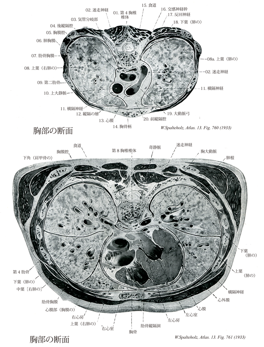
- 760_01【Body of 4th thoracic vertebra; Fourth thoracic vertebral body第4胸椎椎体 Corpus vertebra (Vertebra thoracic IV)】
→()
- 760_02【Vagus nerve [X]迷走神経[脳神経X] Nervus vagus [X]】 Nerve arising from the fourth and fifth pharyngeal arches. It emerges from the medulla oblongata together with CN IX in the posterolateral sulcus and passes through the jugular foramen. Its distribution area extends into the thoracic and abdominal cavities.
→(迷走神経は第10脳神経で、上方の舌咽神経、下方の副神経の間で延髄の外側から多数の小根によって起こる混合神経で胸腹部の諸内臓に分布する副交感神経節前神経線維(延髄迷走神経背側核に細胞体をもつニューロンの神経突起)を主成分としている。これらの線維が胸腹部を走行するあいだに、きわめてしばしば自律神経叢を形成してどこに神経の本幹が存在するか不明瞭となるため、迷走神経の名がつけられた。また迷走神経には胸腹部の内臓の知覚を伝える神経線維(その細胞体は迷走神経の下神経節内に存在する)、咽頭下部および後頭の筋への運動線維(延髄疑核に発し、咽頭に分布するものは舌咽神経からの枝とともに咽頭壁において咽頭神経叢を形成したのち筋に分布する)、咽頭下部および後頭の粘膜への知覚神経線維、などが含まれる。後頭に分布する運動および知覚神経線維は下神経節の直下で後頭に向かう上喉頭神経となるか、あるいは胸腔内で迷走神経本幹から下喉頭神経として分かれて頚部を反回神経として上行するかして目的の器官に達する。)
- 760_03【Tracheal bifurcation気管分岐部 Bifurcatio tracheae】 Asymmetrical bifurcation of the trachea at the level of T4.
→(気管が気管支に分かれる所は第四胸椎の高さに相当し、気管分岐部と呼ばれている。)
- 760_04【Posterior mediastinal space後縦隔腔 Spatium mediastinale posterius; Cavum mediastinale posterius】
→()
- 760_05【Pleural cavity胸膜腔 Cavitas pleuralis; Cavum pleurae】 Slitlike space between the parietal and visceral pleura containing a capillary layer and a small amount of serous fluid.
→(胸膜腔とは、壁側胸膜とその続きである肺胸膜によって囲まれる閉じた空間であって、その内部に存在するのは少量の液(漿液serous fluid)だけである。肺は胸腔の中にあるけれども、胸膜腔の外にある。胸腔の中には心膜によて囲まれる心膜腔という閉じた袋もあり、これらに介在して胸部内臓や大血管などが胸腔内に詰まっているわけである。)
- 760_06【Visceral pleura; Pulmonary pleura臓側胸膜;肺胸膜 Pleura visceralis; Pleura pulmonalis】 Part of the pleura that invests the lung and passes into the interlobar spaces.
→(肺表面を被う胸膜で、肺葉の間にも入り込み、肺の葉間面を被っている。肺胸膜は肺の内側面にある肺門で肺に出入りする肺根を包んだのち、壁側胸膜に移行する。肺間膜では裂溝に沿って陥入している。)
- 760_07【Costal part of parietal pleura; Costal pleura肋骨胸膜;肋骨部(壁側胸膜の) Pars costalis pleurae parietalis; Pleura costalis】 Part of the parietal pleura that lies on the inner surface of the thoracic wall.
→(壁側胸膜の肋骨胸膜は、胸壁の内面(胸骨、肋軟骨、肋骨、肋間筋、肋間膜、胸椎の側面)を覆う。)
- 760_08【Right superior lobe of lung上葉(右肺の) Lobus superior (Pulmo dexter)】
→()
- 760_08a【Superior lobe of lung; Upper lobe of lung上葉(肺の) Lobus superior pulmonis】 Superior lobe that extends posteriorly to the fourth rib. In the right lung, its inferior border runs anteriorly at about the level of the fourth rib. In the left, its inferior border extends to the osseocartilaginous border of the sixth rib.
→(後方では第四肋骨まで達する。右側では、その下端は第四肋骨にほぼ沿って、前方へいたる。左側では、第六肋骨の骨軟骨境界まで達する。 (Feneis))
- 760_09【Second rib [II]第二肋骨 Costa secunda [II]】 Rib that begins at the level of the sternal angle. It is easily palpated.
→(第2肋骨は第1肋骨と第3肋骨との中間形を示している。ただ肋骨溝に相当する溝が上面にも現れる。)
- 760_10【Superior vena cava上大静脈 Vena cava superior; Vena cava cranialis】
→(上大静脈は上半身の血液を集める静脈で、上縦隔の中で左右の腕頭静脈が合してはじまり、途中で奇静脈を受け入れながら上行大動脈の右側を下行して右心房にそそぐ。)
- 760_11【Phrenic nerve横隔神経 Nervus phrenicus】 Nerve arising from C4 with accessory branches from C3 and C5. It lies on the anterior scalene muscle and then passes anterior to the hilum of lung to the diaphragm, with some fibers continuing into the peritoneum.
→(第3~5頚神経から出て頚神経叢を形成し、主に第4頚神経から起こる。頚部では前斜角筋の前面に沿って、また胸腔中では縦隔胸膜と心膜との間を通って、それぞれ走行する。横隔膜にいたる運動神経であるが、壁側縦隔胸膜、心膜、横隔胸膜、腹膜に知覚神経を送り(心臓枝)、腹腔神経叢からの枝と交通する(横隔腹枝)。時に鎖骨下筋神経または腕神経叢の他の神経から小枝が出てて、第1肋骨付近の高さで横隔神経に合することがあるが、これを副横隔神経という。)
- 760_12【Mediastinum縦隔 Mediastinum】 Part of the thorax between the two pleural sacs. It extends from the anterior surface of the vertebral column to the posterior surface of the sternum and from the superior thoracic aperture to the diaphragm. Its connective tissue is continuous with the cervical connective tissue and it communicates with the abdominal cavity via openings in the diaphragm.
→(縦隔は、左右肺の間の部分。矢状方向に、胸骨の後面および肋骨の一部から胸椎の椎体の前面まで広がっている。両側は、縦隔胸膜に接し、下方は横隔膜に境される。縦隔は上縦隔と下縦隔に区分され、下縦隔はさらに後縦隔(脊柱前面と心膜後面との間)、中縦隔(心臓と心膜)、前縦隔(心膜と胸壁の間)に区分されている。)
- 760_13【Pericardium心膜 Pericardium】 Lubricant-containing sheath enclosing the heart. It consists of a fibrous layer and a double-layered serous coat.
→(心膜は心臓と大血管起始部の被覆と活動のための膜。外層の線維性心膜fibrous pericardiumと内層の漿膜性心膜serous pericardiumの2層からなる閉鎖嚢。漿膜性心膜は心臓表面を直接おおう臓側板(心外膜)と線維性心膜の内面をおおう壁側板にわけられる。線維性心膜は強靱な膜で、大血管の壁につづき、心臓を固定・保持するとともに、その急激な過度の拡張を防ぐ。さらに心臓は心膜腔で囲まれ、潤滑な心膜性心膜で包まれるので、摩擦なく拍動することができる。)
- 760_14【Manubrium of sternum胸骨柄 Manubrium sterni】 The part of the sternum above the sternal angle.
→(胸骨柄は胸骨の上1/4を占める部分で、その上縁の左右両端には小刀で角を落としたような切れ込み(鎖骨切痕)があり、ここに鎖骨と連結するための関節面が見える。左右の鎖骨切痕に挟まれた部分の上縁も浅い切れ込みになっている(頚切痕)。また胸骨柄の側面で鎖骨切痕のすぐ下には第1肋骨が接するための切れ込みがある。胸骨柄と胸骨体とが結合する(胸骨剣結合)部位は前方に突出して、後方に開く鈍角すなわち胸骨角を作る。)
- 760_15【Oesophagus; Esophagus食道 Oesophagus; Esophagus】 Passageway measuring 23-26 cm in length that begins below the cricoid cartilage at the level of the sixth cervical vertebra and ends at the cardia of the stomach.
→(食道は咽頭につづき、下方は胃に流入する長い管で、狭義の消化管の最初の部分である。輪状軟骨下縁(上食道狭窄)にはじまり、脊柱の前を下って胃の噴門部に接合するまで、全長23~26cm。内腔は適宜拡がり、義歯を飲み込んだ例もある。内腔の狭い部分は上端(上食道狭窄)、大動脈弓・気管支と交叉する部分(中食道狭窄)、下端(下食道狭窄)の3カ所で、上下端では内腔が普通は閉じ、括約筋の存在が想定されている。食道を上から頚部・胸部・腹部に分ける。頚部は脊椎の前にある部分、胸部は以下横隔膜で、腹部は横隔膜の食道裂孔を抜けて腹腔内に入り、噴門部に流入する短い部分である。食道の壁の粘膜は重層扁平上皮におおわれ、粘膜筋板を有し、食道腺が散在する。上部または下端に食道噴門腺をみる。筋層は上部で横紋筋、下部で平滑筋で、平滑筋束の一部は気管支食道筋、胸膜食道筋として、周囲の器官に連続する。筋層の外側は疎性結合組織性の外膜におおわれる。)
- 760_16【Sympathetic trunk交感神経幹 Truncus sympathicus】 Chain of ganglia that are connected by nerve fibers and lie on the left and right sides of the vertebral column, extending from the base of the cranium to the coccyx.
→(脊椎全長の両脇に1本ずつの交感神経幹(神経節のためのふくらみをそなえる)が存在している。同幹の頚部領域には3個、胸部領域には11~12小、腰部領域には5個、仙骨部領域(骨盤内)には4~5個の幹神経節がある。左右の交感神経幹は脊柱に近接しており、脊柱下端の所では1個の不対神経節につながる。(求心性神経線維) 内臓からの感覚を伝える有髄性の求心性神経線維は交感神経節を素通りして、白交通枝を介して脊髄神経内に入り、その脊髄神経節が所属する髄節の高さの後根神経節の中に含まれる神経細胞体に達する。同じ細胞体からの、中枢に向かう軸索がそののち脊髄に入り、脊髄内での内臓反射路の形成にあずかったり、あるいは脳の自律神経中枢にまで上行したりする。)
- 760_17【Recurrent laryngeal nerve反回神経 Nervus laryngeus recurrens; Nervus recurrens】 Branch of the vagus nerve that extends on the right around the subclavian artery and on the left around the aortic arch. It runs in the groove between the trachea and esophagus to the Larynx. Its terminal portion penetrates the inferior pharyngeal constrictor and supplies the mucosa to about the rima glottis as well as all laryngeal muscles with the exception of the cricothyroid. It communicates with the internal branch of superior laryngeal nerve.
→(右側は鎖骨下動脈をまわり、左側では大動脈弓を回って気管と食道の間の溝にはいる。そして気管に気管枝を、食道に食道枝をそれぞれ送る。さらに終枝として下喉頭神経を、下咽頭収縮筋を貫いて輪状筋以外の喉頭筋と喉頭下半分の粘膜に送る。)
- 760_18【Inferior lobe; Lower lobe of lung下葉(肺の) Lobus inferior pulmonis】 It mainly extends dorsally. Its superior border runs obliquely from posterosuperior to anteroinferior. It begins paravertebrally at the fourth rib and ends at the intersection of the middavicular line and the sixth rib.
→(後側に主な拡がりをもつ。その上限界上後方から下前方へ斜めに走り、第四肋骨から脊柱側方で鎖骨中間線を第六肋骨が切る線までいたる。 (Feneis))
- 760_19【Arch of aorta; Aortic arch大動脈弓 Arcus aortae】 It is located between the ascending and descending aorta. Its roof extends to the first rib at the left border of the sternum.
→(大動脈弓は上行大動脈につづく弯曲部であり(約5~6cm長)、肺動脈分岐部および左気管支をこえて左後方にまわり、第四胸椎体の左側で胸大動脈に移行する。)
- 760_20【Anterior mediastinal space前縦隔腔 Spatium mediastinale anterius; Cavum mediastinale anterius】
→()