Spalteholz HANDATLAS DER ANATOMIE DES MENSCHEN VON WERNER SPALTEHOLZ
メニューは解剖学(TA)にリンクしてあります。図の番号をクリックすると下記の説明へ、右側の用語をクリックすると解剖学(TA)にジャンプします。
965


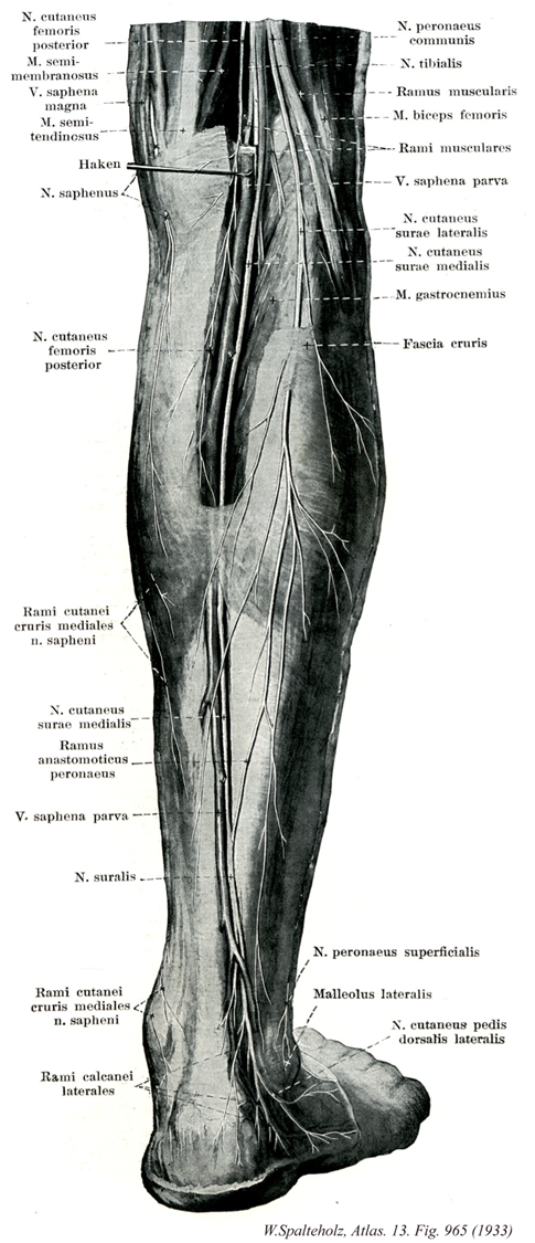
- 965_00【Leg region下腿部 Regio cruris】
→(下腿部は脛骨と腓骨。大腿と同様に前面の前下腿部と後面の後下腿部および内側(脛側)面、外側(腓骨)面とに分けられる。)
- 965_01【Posterior cutaneous nerve of thigh; Posterior femoral cutaneous nerve後大腿皮神経;後皮神経(大腿の) Nervus cutaneus femoris posterior】 Nerve arising from SI-S3 that travels through the greater sciatic foramen distal to the piriformis and supplies the skin on the posterior side of the thigh as well as the proximal part of the leg.
→(後大腿皮神経は仙骨神経叢のS1~S3より起こる。大坐骨孔を通り梨状筋の下で臀部にあらわれる。この神経は坐骨神経と大臀筋ではさまれながら臀部を下行し、大腿二頭筋よりも浅層を走りながらこれを横切り大腿背面の深筋膜内を走る。膝窩に達してから後大腿皮神経本幹は深筋膜を貫き皮膚に向かう。)
- 965_02【Semimembranosus muscle半膜様筋 Musculus semimembranosus】 o: Ischial tuberosity. i: Medial condyle of tibia and oblique popliteal ligament. It is partly covered by the semitendinosus muscle. Extension at the hip joint; flexion and medial rotation at the knee joint. Tenses the knee joint capsule. I: Tibial nerve.
→(半膜様筋は大腿二頭筋長頭と大腿方形筋の起始の間の坐骨結節から起こる。脛骨内側顆、膝関節包後壁および膝窩筋の筋膜に停止する。半膜様筋は中4分の2のみが筋性である。起始腱は広い腱性の板をなし、停止腱も同じ平板である。3本の腱様の索として終わる。脛骨への索は腹側で迂回し、内側側副靱帯の下の脛骨内側顆に付く。中央の索は筋の方向を受け継ぎ、一部は脛骨近位端後面に、一部は膝窩筋の筋膜に付く。腓骨への索は膝関節包の後壁を補強し、斜膝窩靱帯として大腿骨外側顆に向かって外側へ射創する滑液包が通常同筋の停止腱と脛骨内側顆の間にある。)
- 965_03【Great saphenous vein; Long saphenous vein大伏在静脈 Vena saphena magna】 Vein possessing valves that arises on the medial side of the foot and ascends medially, collecting most of the medial superficial cutaneous veins. It passes through the saphenous opening to empty into the femoral vein.
→(大伏在静脈は古くは薔薇静脈とよんだこともある。ギリシャ語のsaphisは「見える」という意味であるというが、他方アラビア語では「かくれた」という意味を表すという。このように語原的にはギリシャ語とアラビア語では反対の意味に解されているのは興味深いが、いずれにしてもVena saphenaの語原ははっきりしていない。大伏在静脈は下肢最大の皮静脈で、下肢の内側に沿って皮下組織の中を上行する。足背の内側縁にはじまり、内果の前を通るが、この部分で皮膚のうえからその走行をみることができる。伏在神経と伴行して下腿の内側を通り、膝関節の後内側を経て大腿内側面を上行し、鼡径靱帯の下方で深く入り、伏在裂孔で大腿静脈にそそぐ。この経過の途中、周辺より多くの皮静脈がこれに合流するが、とくに大腿の内側と後面よりの皮静脈は1本に合して伏在裂孔のやや下方で大伏在静脈にそそぐことがある。これを副伏在静脈という。また大腿の前面や外側面よりの皮静脈が合しこれにそそぐときは、とくに外側伏在静脈とよぶことがある。このときは大内々側面よりのものは内側伏在静脈という。)
- 965_04【Semitendinosus muscle半腱様筋 Musculus semitendinosus】 o: Ischial tuberosity. i: Medial surface of tibia. Extension at the hip joint. Flexion and medial rotation at the knee joint. I: Tibial nerve.
→(半腱様筋は大腿二頭筋長頭の起始近くの坐骨結節から起こり、鵞足を介して脛骨近位端内側面および下腿筋膜に終わる。半腱様筋は半膜様筋によってつくられた溝の中を遠位へ向かう。長い停止腱は大腿部ですでに始まり(ここから“半腱様”の名がつけられた)、鵞足の深層へと放散する。)
- 965_05【Saphenous nerve伏在神経 Nervus saphenus】 Longest, purely sensory branch of the femoral nerve. It begins in the femoral triangle, passes beneath the 「vastoadductor membrane,」 which it pierces, continues between the sartorius and gracilis to beneath the skin, and then travels with the great saphenous vein as far as the medial side of the foot.
→(大腿三角から足に至る大腿神経の枝。伏在神経は大腿動脈の外側を沿って走り、動脈とともに内転筋管内を下降する。膝関節の内側で皮下にでて大伏在静脈に沿って下行し、下腿と足背との内側面の皮膚に分布する。)
- 965_06【Medial cutaneous nerve of leg; Medial crural cutaneous nerve内側下腿皮枝;脛側下腿皮枝(伏在神経の) Rami cutanei cruris mediales (Nervus saphenus)】 Branches of the saphenous nerve that extend to the skin of the leg and foot.
→(伏在神経の主幹より起こり、下腿および足の内側面の皮膚へ分布。 (Feneis))
- 965_07【Medial sural cutaneous nerve内側腓腹皮神経 Nervus cutaneus surae medialis】 Nerve given off by the tibial nerve in the popliteal fossa that descends subfascially lateral to the small saphenous vein and joins the sural communicating branch to form the sural nerve.
→(脛骨神経より起こり、膝窩へ入り、小伏在静脈の外側で筋膜下を下行する。腓腹神経との交通枝と合し、腓腹神経となる。 (Feneis))
- 965_08【Sural communicating branch of lateral sural cutaneous nerve腓腹交通枝;腓側交通枝(外側腓腹皮神経の) Ramus communicans fibularis; Ramus communicans peroneus; Ramus communicans nervus peronei communis cum nervus cutaneo surae mediali fibularis; Ramus communicans nervus fibularis communis cum nervus cutaneo surae medialis】 Branch that travels deep to the fascia over the lateral head of gastrocnemius. It joins the medial sural cutaneous nerve to form the sural nerve.
→(外側腓腹皮神経の腓腹神経との交通枝は総腓骨神経の腓腹神経との交通枝。膝窩内の総腓骨神経から起こり、腓腹筋の外側頭上を越えて下腿の中1/3に達し、内側被覆皮神経と結合して腓腹神経を形成する。)
- 965_09【Small saphenous vein; Short saphenous vein小伏在静脈 Vena saphena parva】 Vein from the lateral border of the foot that passes over the posterior aspect of the leg to the popliteal vein.
→(小伏在静脈は足背の外側縁にはじまり、外果の後面を通って踵骨腱の外側に沿って上行し、次いで下腿後面のほぼ中央で下腿筋膜の表層を通り、膝窩の下部で筋膜を貫いて深層に入り、腓腹筋の両頭の間から膝窩静脈にそそぐ。皮下組織の中を走る皮静脈で、その経過中、腓腹神経と伴行する。)
- 965_10【Sural nerve腓腹神経 Nervus suralis】 Continuation of the medial sural cutaneous nerve after it joins the sural communicating branch.
→(腓腹神経は脛骨神経の皮枝で腓腹筋の2頭のあいだを下行するが、その途中で総腓骨神経からの交通枝を受けるのがふつうである。腓腹神経からの小枝が下腿後面の皮膚に分布する。外顆より遠位では腓腹神経は小伏在静脈と伴行しながら、足の外側縁から小指外側縁に至る皮膚に枝を起こる。 (Netter)腓腹神経は皮神経であり、膝窩の中部あるいは下部で脛骨神経より起こる(図13)。腓腹筋の両頭の間を下行しながら、腓腹神経は下腿筋膜を貫通して小さな内側腓腹皮神経(内側腓腹皮神経は下腿筋膜貫通して小さな内側腓腹皮神経(内側腓腹皮神経はもっとも大きく、脛骨神経より直接起こることもある)を出し、外側腓腹皮神経から腓腹神経との交通枝を受ける。腓腹神経は、小伏在静脈近傍を通ってアキレス腱の外側へと下行を続けながら、下腿背側面および外側面を覆う皮膚と筋膜に枝を与える。外果と踵骨腱(アキレス腱)の間に達すると腓腹神経は外側踵骨枝を出し、足根と踵の外側面の皮膚と筋膜枝を出す(これに対応する内側踵骨枝は脛骨神経から起こる)。腓腹神経の終末枝は、外側足背皮神経として足および小趾の外側に沿って前方へ走る。)
- 965_11【Lateral calcaneal branches of sural nerve外側踵骨枝;腓側踵骨枝(腓腹神経の) Rami calcanei laterales (Nervus suralis)】 Lateral branches to the heel.
→(腓腹神経の外側踵骨枝は踵骨へ向かう腓腹神経の外側枝である。)
- 965_12【Common fibular nerve; Common peroneal nerve総腓骨神経 Nervus fibularis communis; Nervus peroneus communis】 Branch of the sciatic nerve arising from L4-S2. It accompanies the tendon of the biceps femoris to posterior to the head of fibula and then crosses obliquely forward between the skin and the fibula.
→(総腓骨神経はL4~S2より起こる。坐骨神経の2終枝のうちの細い方である。大腿の下1/3の高さから始まり、膝窩内を下腿への皮神経を出しながら、大腿二頭筋の停止腱の後内側縁に沿って腓骨頭の後面に達し、ここで下腿筋の下層にもぐり込む。腓腹筋外側頭よりも浅層でこれを横切り、膝窩を離れる。以上の経過中に総腓骨神経は腓骨頚外側面で皮膚のみに被われた状態となるため、体表よりこの神経を容易に触知できる。そののち総腓骨神経は腓骨頭の後方をやや下行してから急に向きを変えて長腓骨筋内に侵入し、そこで浅腓骨神経および深腓骨神経に分かれる。)
- 965_13【Tibial nerve脛骨神経 Nervus tibialis】 Second terminal branch of the sciatic nerve arising from L4-S3. It travels through the popliteal fossa, passes deep to the tendinous arch of the soleus, and accompanies the posterior tibial artery around the medial malleolus to the sole of the foot.
→(脛骨神経はL4~S3より起こる。坐骨神経の第二の終枝。膝窩を通りヒラメ筋腱弓の下をすぎ後脛骨筋とともに内果をまわり、足底へ達す。下腿の屈筋群、足底の諸筋、下腿の後面と足底の皮膚に分布するが、次の神経はいずれも脛骨神経の末梢枝である。①下腿骨間神経(下腿骨間膜の後縁に沿って走り、足関節のあたりに達する)、②内側被覆皮神経、腓腹神経、外側足背神経(ひとつづきのもので下腿後面から足背外側部の皮膚に分布)、③内側足底神経と外側足底神経(ともに足底の諸筋に分布する枝を出したあと、趾の足底面や足底の皮膚に分布するため、総底側趾神経に枝分かれし、固有底側趾神経となっておわる)。)
- 965_14【Muscular branches of common fibular nerve筋枝(総腓骨神経の) Rami musculares (Nervus fibularis communis)】 Branches to the peroneus longus and brevis.
→(浅腓骨神経の筋枝は長および短腓骨筋への枝。)
- 965_15【Biceps femoris muscle大腿二頭筋 Musculus biceps femoris】 Two-headed muscle arising from the pelvis and femur, i: Head of fibula. Flexion at the knee joint, lateral rotation.
→(大腿二頭筋は2関節性の長頭と1関節性の短頭から成る。長頭は坐骨結節で半腱様筋と総頭をつくって起こる。短頭は粗線の外側唇の中1/3と外側筋間中隔から起こる。これら両頭は合して2頭筋となり、腓骨頭に終わる。その際この筋と膝関節の外側側副靱帯との間に大腿二頭筋の下腱下包がある。股関節では長頭は大腿を後斜するように働く。膝関節では大腿二頭筋は屈曲するように働き、屈曲した状態では下腿を外旋する。この筋は膝関節における唯一の外旋筋であって、すべての内旋筋に匹敵する作用をもっている。)
- 965_16【Muscular branches of tibial nerve筋枝(脛骨神経の) Rami musculares nervus fibularis profundus; Rami musculares nervus tibialis】 Branches to the gastrocnemius, plantaris, soleus, and deep flexors of the leg.
→(脛骨神経の筋枝は腓腹筋の2頭、足底筋、ヒラメ筋、膝窩筋に侵入する。)
- 965_17【Lateral sural cutaneous nerve外側腓腹皮神経;腓側腓腹皮神経 Nervus cutaneus surae lateralis; Nervus cutaneus surae fibularis】 Nerve given off in the popliteal fossa that supplies the skin of the lateral side of the leg as well as the upper two-thirds of its posterior side.
→(外側腓腹皮神経は膝窩よりあらわれ、たいていは下腿の後外側面の上2/3の皮膚へ分布する。)
- 965_18【Gastrocnemius muscle腓腹筋 Musculus gastrocnemius】 Superficial leg muscle composed of the following two heads. Flexion at the knee joint, plantar flexion and supination at the ankle joint.
→(下腿後側の浅層の筋で、ヒラメ筋と合わせて下腿三頭筋と呼ばれる。大腿骨の内側上顆(内側頭)と外側上顆(外側頭)とから起こる。その停止腱はヒラメ筋とともに合流し、アキレス腱(踵骨腱)となり、踵骨隆起に停止する。踵骨後面の上部と踵骨腱が接近する部位には小さな滑液包が介在する。この筋は脛骨神経支配を受け、距腿関節による足の底屈、膝関節屈曲を生じさせる。Gastrocnemiusはフクラハギ(gastro-腹[腹のようにふくらむ]+kneme脚、スネ))
- 965_19【Fascia lata大腿筋膜 Fascia lata】 Fascia enclosing the thigh muscles. It is attached anteriorly to the iliac crest and inguinal ligament, splitting into two layers medial to the sartorius muscle and deep to the inguinal ligament. It forms the C-shaped lateral margin of the saphenous opening and covers the femoral vessels with a perforated sievelike sheet. Its deep portion lies posterior to the femoral vessels. Both layers unite with the pectineal fascia. Laterally it thickens to form a tendinous band. It is continuous with the gluteal fascia to posterosuperior and with the deep fascia of the leg to distal.
→(大腿筋膜は大腿筋全体の表面を包み、上方は鼡径靱帯、腸骨稜、仙骨外側縁および恥骨弓についてから浅腹筋膜および浅背筋膜に連なり、下方は下腿筋膜につづく。よく発達して厚いが、とくに外側部は腱膜様に著しく厚くなって腸脛靱帯となっている。)
- 965_20【Superficial fibular nerve; Superficial peroneal nerve浅腓骨神経 Nervus fibularis superficialis; Nervus peroneus superficialis】 Terminal branch of the common fibular nerve. It descends between the peroneus muscles and extensor digitorum longus.
→(浅腓骨神経は総腓骨神経の終枝の一つ、腓骨筋と長趾伸筋の間を下行する。 (Netter)浅腓骨神経は、長趾伸筋と腓骨筋の間を下行し、長腓骨筋と短腓骨筋に筋枝を出した後、下腿の虫部から下部1/3に移る高さで下腿筋膜を貫く。この高さで、浅腓骨神経は、内側足背皮神経と中間足背神経とに分かれる。内側足背皮神経は足根の前面を走行して足背に至り、下部下腿前面と足背の皮膚と筋膜に枝を送る。下伸筋支帯の下縁近くで、この神経は2本の足背趾神経に分岐する。このうち1本は、足背および母趾の内側面と背側面を支配し、他の1本は第2,第3趾の背側面と側面とを支配する。中間足背皮神経は、足背外側部に沿って走行し、近傍の皮膚や筋膜に枝を出し、第3と第4趾および第4と第5趾に行く2本の足背趾神経に分かれる。また、中間足背皮神経は、外側足背皮神経と交通する。)
- 965_21【Lateral malleolus外果;外踝;腓骨踝 Malleolus lateralis; Malleolus fibulae】
→(腓骨の下端は肥厚して下方に突出し、特にその外側面を外果とよぶ。外果の先端が下方というよりは後下方を向いている。臨床的には、腓骨の骨折はしばしば見られ、スキー人口の増加とともに外果の骨折が激増している。これはスキーで転んで足首のところで足が強く内反されると、外果窩についている強い靱帯が引っ張られて、外果の先端から約1cm上方の所で外果が簡単に折れてしまう。)
- 965_22【Lateral dorsal cutaneous nerve外側足背皮神経;腓側足背皮神経 Nervus cutaneus dorsalis lateralis; Nervus cutaneus dorsi pedis fibularis】 Branch to the lateral portion of the dorsum of foot. It anastomoses with the intermediate dorsal cutaneous nerve.
→(外側足背皮神経は腓腹神経の延長で、足背の外側縁とその付近を支配する部分である。)