Spalteholz HANDATLAS DER ANATOMIE DES MENSCHEN VON WERNER SPALTEHOLZ
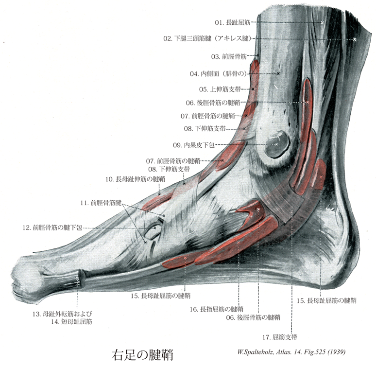
メニューは解剖学(TA)にリンクしてあります。図の番号をクリックすると下記の説明へ、右側の用語をクリックすると解剖学(TA)にジャンプします。
525 


- 525_01【Flexor digitorum longus muscle長趾屈筋;長指屈筋(足の) Musculus flexor digitorum longus】 o: Tibia, i: Distal phalanges of the second through fifth toes. Plantar flexion, supination, flexion of toes. I: Tibial nerve.
→(長趾屈筋はヒラメ筋線より遠位の脛骨後面およびヒラメ筋腱弓の一部から起こる。その停止腱は基節骨の領域で短趾屈筋の腱を貫通し(第2~5)趾の末節骨に停止する。長趾屈筋の腱は腱鞘に包まれて、内果溝を後脛骨筋腱の背外側に走り、屈筋死体の下を載距突起内側縁に沿って足底に至る。舟状骨粗面のレベルでは長母趾屈筋腱の浅層を通る。この際、長母趾屈筋の健束が長趾屈筋の腱に混じる。この腱交叉位遠では足底方形筋が長趾屈筋の腱に付く。この付加的な屈筋は長趾屈筋停止腱の牽引方向を趾放線の長軸方向と関連させる。同一趾へ向かう長趾屈筋(「貫通筋」と短趾屈筋「被貫通筋」)の停止腱は腱鞘(滑液鞘に包まれる。腱鞘は第1中足骨頭の上方から始まり、末節骨までのびている。これらの滑液鞘は線維鞘に包まれる。線維鞘は手指におけると同じように横走線維と交織する線維(輪状および十字部)からなる。)
- 525_02Achilles tendon【Calcaneal tendon踵骨腱;下腿三頭筋腱;アキレス腱 Tendo calcaneus; Tendo musculus tricipitis surae】 Tendon of the triceps surae that attaches on the calcaneal tuberosity.
→(アキレス腱とも呼ばれる。踵骨腱(下腿三頭筋の停止腱)。ギリシャの英雄アキレスの唯一の弱点が踵にあったことから名づけられた。アキレスとはホーマーの叙事詩イリアドIliadの主人公でギリシャの英雄である。彼が将来トロイとの戦いで戦死するだろうという予言を聞いた母のテーティスThetis(海の女神)は、赤ん坊のアキレスをスティックス河(冥界の川)の聖流に浸して不死身のからだとした。けれどもそのとき母親がアキレスの足首をつかんでいたので、踵の所だけが魔法の水にぬれず、彼の唯一の泣きどころになってしまった。立派に成人したアキレスは、トロイ戦争でギリシャ軍に加わって、数々の武功を立てたが、トロイの応じパリスParisが放った矢がアキレスの踵を射抜き、さすがの彼も倒れたというのである。降って1693年にベルギーの解剖学者P.Verheyenが切断された自分の足を解剖しながら、イリアドの故事を思い起こして、踵骨腱のことを初めてアキレス腱と名付けたといわれる。)
- 525_03【Tibialis anterior muscle前脛骨筋 Musculus tibialis anterior】 o:Lateral surface of tibia, interosseous membrane, deep fascia of leg. i: Medial aspects of medial cuneiform and first metatarsal. Dorsiflexion and supination of foot. I: Deep fibular nerve.
→(前脛骨筋は脛骨外側顆、脛骨外側面(近位2/3)、下腿筋膜および筋間膜から起始する。第1中足骨と第1楔状骨あたりの足底部に停止する。収縮中に筋腹は脛骨近位1/3の骨縁上に突出する。その腱は脛骨遠位1/3にかけて形成され、伸筋支帯の下を通って足の内側縁へ至る。その腱鞘は伸筋支帯より近位に始まり、距腿関節の関節腔のレベルにまで伸びている。腱鞘は前脛骨筋腱の遠位部および近位部浅層をおおい、中間部を包んでいる。前脛骨筋と長趾伸筋に対する近位の筋枝は深腓骨神経から同神経がまだ腓骨筋群を容れる部位を通っている内に分かれる。深腓骨神経が長趾伸筋を貫通してから遠位の筋枝が両筋の各々に行き(通常2条の)筋枝が母趾の伸筋へ行く。)
- 525_04【Medial surface of fibula内側面;脛側面(腓骨の) Facies medialis; Facies tibialis】 Surface of the shaft facing the tibia between the anterior and interosseous borders.
→(腓骨の内側面はややくぼんだ、はなはだ幅が狭い面で、骨間膜と前下腿筋間中隔とにはさまれた下腿伸筋群が起こる場所となる。)
- 525_05【Superior extensor retinaculum of foot上伸筋支帯[足の];下腿横靱帯 Retinaculum musculorum extensorum superius pedis; Ligamentum transversum cruris】 Transverse thickening of the deep fascia of the leg that is about two fingers' width and holds the extensor tendons in place.
→(足の上伸筋支帯(下腿横靱帯lig. Transversum cruris)は下腿筋膜の下部が厚くなったもので、伸筋の筋と腱の移行部を被って内果と外果のやや上方で脛骨と腓骨につき、後方は深下腿筋膜に移行する。)
- 525_06【Tendinous sheath of tibialis posterior後脛骨筋の腱鞘 Vagina tendinis musculi tibialis posterioris】 Tendon sheath surrounding the tibialis posterior beneath the flexor retinaculum. It begins where it is crossed by the flexor digitorum longus.
→(後脛骨筋の腱鞘は屈筋支帯に被われて、後脛骨筋の腱の周囲をかこみ、さらに遠位側にのびる。)
- 525_07【Tendinous sheath of tibialis anterior前脛骨筋の腱鞘 Vagina tendinis musculi tibialis anterior】 Tendon sheath of the tibialis anterior that already begins beneath the extensor retinaculum.
→(前脛骨筋腱鞘は伸筋支帯の深部にある滑膜性腱鞘で、足関節前部を通るとき前脛骨筋の腱を囲む。)
- 525_08【Inferior extensor retinaculum of foot下伸筋支帯[足の];下腿十字靱帯 Retinaculum musculorum extensorum inferius pedis; Ligamentum cruciforme cruris】 Thickened portions of the deep fascia of the leg that extend as cruciate bands from both malleoli to the opposite margins of the foot.
→(足の下伸筋支帯は足背筋膜の近位部が厚くなったもので、踵骨外側面の前上部から起こって内果と内側楔状骨に向かうY字形をなす。下伸筋支帯の外側脚は最も強く、第3脛骨筋、長趾伸筋の腱および短趾伸筋の浅深両面を包む。これをワナ靱帯(INA)ともいう。上内側脚はやや弱く、長母趾伸筋の浅面と前脛骨筋の唇面を通り内果につくが、さらに弱い層がこの2腱の他の面を包む。下内側脚は最も弱く長母趾伸筋の浅面を被う、そのほか下内側脚のさらに前方長・短母趾伸筋を被うものがある(母趾伸筋支帯)。またまた外側脚のほかに上外側脚があるときは、外果から起こり、全体として十字状となる。)
- 525_09【Subcutaneous bursa of medial malleolus内果皮下包;脛骨踝皮下包 Bursa subcutanea malleoli medialis】 Bursa situated between the skin and the medial malleolus.
→(内果皮下包は脛骨内果の皮下にある。)
- 525_10【Tendinous sheath of extensor hallucis longus長母趾伸筋の腱鞘;長母指伸筋腱鞘(足の) Vagina tendinis musculi extensoris hallucis longi】 Sheath surrounding the long extensor tendon of the great toe beneath the extensor retinaculum and further to distal.
→(長母趾伸筋の腱鞘は下伸筋支帯の下から起こり、さらに遠位側にのびる。第3脛骨の腱も共同につつむ。)
- 525_11【Tibialis anterior tendon前脛骨筋腱 Tendo musculus tibialis anterior】
→()
- 525_12【Subtendinous bursa of tibialis anterior前脛骨筋の腱下包;前脛骨筋腱包 Bursa subtendinea musculi tibialis anterior】 Bursa situated between the tendon and the medial cuneiform.
→(前脛骨筋の腱下包の前脛骨筋の腱と内側楔状骨または第1中足骨との間にある。)
- 525_13【Abductor hallucis muscle母趾外転筋;母指外転筋(足の) Musculus abductor hallucis】 o: Calcaneal tuberosity. i: Medial sesamoid bone and proximal phalanx of great toe. Medial abduction, supports longitudinal arch of foot. I: Medial plantar nerve.
→(母趾外転筋は踵骨隆起の内側突起、屈筋支帯および足底腱膜から起始する。腱となり内側種子骨を介して母趾の基節骨底内側面および短母趾屈筋の内側腱に停止する。内側足底神経の支配を受ける。この筋の収縮は母趾の屈筋と外転とをもたらす(体重を支えていない下肢の場合)。また、体重を支えている下肢においては、この筋の収縮が内側縦足弓の維持に役立つ。 )
- 525_14【Flexor hallucis brevis muscle短母趾屈筋;短母指屈筋(足の) Musculus flexor hallucis brevis】 o: Cuneiform (I), long plantar ligament, tendon of tibialis posterior, plantar aponeurosis. Forms the groove for the flexor hallucis longus. Plantar flexion of great toe. I: Medial plantar nerve.
→(短母趾屈筋は楔状骨、底側踵立方靱帯および後脛骨筋の腱から起始する。その内側頭は母趾外転筋の腱とともに内側種子骨を介して中足指節関節に停止する。その外側頭は母趾内転筋の腱とともに外側種子骨を介して基節骨に停止する。短母趾屈筋は内側足底神経の支配を受ける。この筋の収縮により第1趾の中足趾節関節における屈曲が得られる。また、この筋は内側縦束裂を維持する役割も果たす。)
- 525_15【Tendinous sheath of flexor hallucis longus長母趾屈筋の腱鞘;長母指屈筋の腱鞘(足の) Vagina tendinum musculi flexoris hallucis longi】 Tendon sheath surrounding the long flexor tendon of the great toe, extending to the proximal end of the sole of the foot, where it crosses under the tendon of the flexor digitorum longus.
→(長母趾屈筋の腱鞘は下伸筋支帯の下にあって、そこからさらに下方にのびる。)
- 525_16【Tendinous sheath of flexor digitorum longus長指屈筋の腱鞘(足の);長趾屈筋の腱鞘 Vagina tendinum musculi flexoris digitorum longi】 Tendon sheath surrounding the long flexors of the toes, located posterior and inferior to the medial malleolus and covered by the flexor retinaculum.
→(長趾屈筋の腱鞘は足根部内側で長趾屈筋の腱をおおい、屈筋支帯の深部で足に達する滑膜性腱鞘。)
- 525_17【Flexor retinaculum of foot屈筋支帯[足の];破裂靱帯 Retinaculum musculorum flexorum pedis; Ligamentum laciniatum】 Multilayered band situated over the long flexor tendons passing from the medial malleolus to the calcaneus. Its superficial portion invests the tibial nerve and posterior tibial artery and veins. Its deep portion forms an osteofascial canal with compartments containing the posterior tibial flexor muscles, flexor digitorum longus, and flexor hallucis longus.
→(足の屈筋支帯は下腿筋膜の厚くなったもので、内果の下部から扇状に広がって、前部は舟状骨に後部は踵骨につき中間部は足底腱膜に移行する。屈筋支帯は後脛骨筋と長趾屈筋の腱を被い、またその間の隔壁を骨に送ったのち、載距突起についてさらに長母指屈筋腱溝を被う深葉と、脛骨神経および後脛骨動静脈を被う浅葉とに分かれる。)
