Spalteholz HANDATLAS DER ANATOMIE DES MENSCHEN VON WERNER SPALTEHOLZ
メニューは解剖学(TA)にリンクしてあります。図の番号をクリックすると下記の説明へ、右側の用語をクリックすると解剖学(TA)にジャンプします。
573 


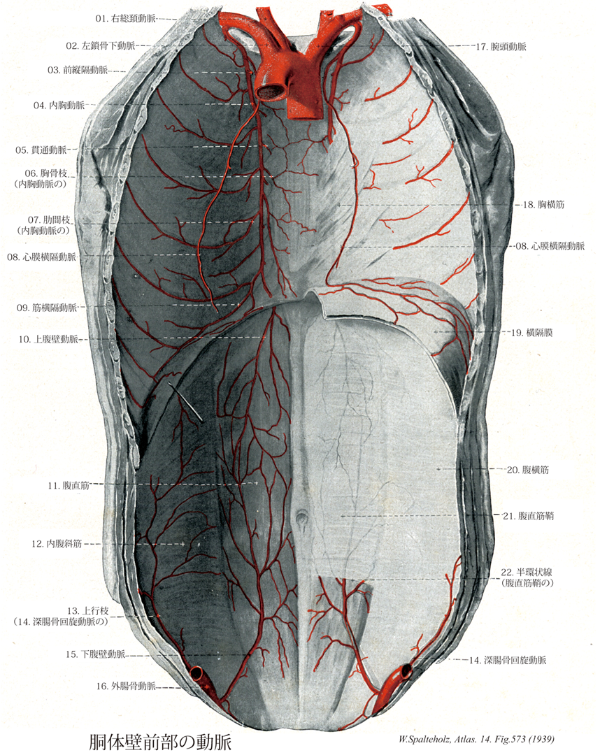
- 573_01【Right common carotid artery右総頚動脈 Arteria carotis communis dextra】
→(総頚動脈は頭部に血液を送る血管の主幹。右は腕頭動脈の枝、左は大動脈弓の上行部より出る。そのため左総頚動脈は右のものよりも4~5cm長い。総頚動脈は枝を出さず、気管・喉頭の両側を上行し、甲状軟骨上縁の高さで音叉のような形をなし内・外頚動脈に分かれる。分岐部の後側には頚動脈小体が存在する。また分岐部のないし内頚動脈始部の壁はやや薄く膨隆しており(頚動脈洞)、舌咽神経の枝を介し血圧を感受するという。)
- 573_01a【Common carotid artery総頚動脈 Arteria carotis communis】 Artery of the neck without any branches. It runs on both sides of the trachea and larynx and passes deep to the sternocleidomastoid. It arises on the right from the brachiocephalic trunk and on the left from the aortic arch.
→(総頚動脈は頭部に血液を送る血管の主幹。右は腕頭動脈の枝、左は大動脈弓の上行部より出る。そのため左総頚動脈は右のものよりも4~5cm長い。総頚動脈は枝を出さず、気管・喉頭の両側を上行し、甲状軟骨上縁の高さで音叉のような形をなし内・外頚動脈に分かれる。分岐部の後側には頚動脈小体が存在する。また分岐部のないし内頚動脈始部の壁はやや薄く膨隆しており(頚動脈洞)、舌咽神経の枝を介し血圧を感受するという。)
- 573_02【Left subclavian artery左鎖骨下動脈 Arteria subclavia sinistra】
→(鎖骨下動脈は上肢の主幹動脈の根部をなし、右側は腕頭動脈から、左側は大動脈弓からそれぞれ分かれてはじまり、前斜角筋の後方を通って第1肋骨外側縁で腋窩動脈につづく。胸・頚・上肢移行部の動脈として、多彩な分枝と変異に富むことを特徴とする。分枝はつぎの通りである。椎骨動脈、内胸動脈、甲状頚動脈、肋頚動脈、下行肩甲動脈に分枝し、第一肋骨を越えたところで腋窩動脈となる。)
- 573_02a【Subclavian artery鎖骨下動脈 Arteria subclavia】 Artery that passes with the roots of brachial plexus between the anterior and middle scalene muscles through the scalene space, over the first rib in the groove for the subclavian artery. From the lateral border of the first rib, it continues as the axillary artery.
→(鎖骨下動脈は上肢の主幹動脈の根部をなし、右側は腕頭動脈から、左側は大動脈弓からそれぞれ分かれてはじまり、前斜角筋の後方を通って第1肋骨外側縁で腋窩動脈につづく。胸・頚・上肢移行部の動脈として、多彩な分枝と変異に富むことを特徴とする。分枝はつぎの通りである。椎骨動脈、内胸動脈、甲状頚動脈、肋頚動脈、下行肩甲動脈に分枝し、第一肋骨を越えたところで腋窩動脈となる。)
- 573_03【Mediastinal branches of internal thoracic artery縦隔枝;前縦隔動脈(内胸動脈の) Rami mediastinales arteria thoracicae internae; Arteriae mediastinales ventrales】 Branches supplying the mediastinum.
→(内胸動脈の縦隔枝は前縦隔にある構造、主に胸腺とリンパ節に分布する小枝。)
- 573_04【Internal thoracic artery内胸動脈 Arteria thoracica interna】 Artery arising from the subclavian artery and descending along the anterior inner side of the thorax to the diaphragm.
→(内胸動脈は胸骨縁に沿い前胸壁内面を下行し、横隔膜前端を貫いて上腹壁動脈に移行し、腹直筋内で下腹壁動脈と吻合して、前正中線に沿う縦走動脈路を形成する。異常の経過からして縦隔と前胸壁に分布するのに適している。縦隔への枝としては、縦隔枝、胸腺枝、気管支枝さらに横隔神経に伴走する心膜横隔動脈がある。前胸壁への枝としては、胸骨枝、肋間隙を外側に走り肋間動脈と吻合する前肋間枝、肋間隙を貫き乳腺枝を分岐しうる貫通枝、ならびに横隔膜と胸壁下部に分布する筋横隔動脈などがある。なお側胸壁内面を下行する外側肋骨枝がまれに内胸動脈初部からおこることがある。)
- 573_05【Perforating branches of internal thoracic artery貫通枝(内胸動脈の);貫通動脈 Rami perforantes (Arteria thoracica interna); Arteria perforantes】 Vessels that penetrate the first through sixth intercostal spaces, passing to the surface of the thorax.
→(内胸動脈の貫通枝は内胸動脈から第1~6肋軟骨間隙を貫いて皮膚および皮下組織に分布する小枝。)
- 573_06【Sternal branches of internal thoracic artery胸骨枝(内胸動脈の) Rami sternales (Arteria thoracica interna)】 Branches to the sternum.
→(内胸動脈の胸骨枝は内側に出て胸横筋や胸骨後面に分布する。)
- 573_07【Anterior intercostal branches of internal thoracic artery; Anterior intercostal arteries前肋間枝;肋間枝(内胸動脈の);前肋間動脈 Rami intercostales anterior; Rami intercostales; Arteriae intercostales anterior】 Anterior tributaries passing into the intercostal spaces.
→(内胸動脈の前肋間枝は胸壁の肋間隙の前部に分布する動脈で、1-6は内胸動脈の枝。7-11は筋横隔動脈の枝である。)
- 573_08【Pericardiacophrenic artery; Anterior bronchial artery心膜横隔動脈;前気管支動脈 Arteria pericardiacophrenica】 It accompanies the phrenic nerve and supplies the pericardium and diaphragm.
→(心膜横隔動脈は内胸動脈より起こり、心膜、横隔膜、胸膜に分布する。筋横隔動脈、下横隔動脈、内胸動脈の縦隔枝・心膜枝と吻合する。)
- 573_09【Musculophrenic artery筋横隔動脈 Arteria musculophrenica】 Artery running posterior to the costal arch and giving off the remaining anterior intercostal branches from the seventh intercostal space onward.
→(筋横隔動脈は肋骨弓の後面を外下方に進み、胸壁の外側下部と横隔膜とに分布する。)
- 573_10【Superior epigastric artery上腹壁動脈 Arteria epigastrica superior】 Continuation of the internal thoracic artery after it enters the abdominal cavity between the sternal and costal parts of diaphragm (Larrey's cleft = sternocostal triangle).
→(上腹壁動脈は内胸動脈の内側終枝より起こり、腹部筋と皮膚、肝鎌状間膜に分布する。下腹壁動脈と吻合する。)
- 573_11【Rectus abdominis muscle腹直筋 Musculus rectus abdominis】 o: Fifth to seventh costal cartilages, xiphoid process, i: Pubic crest and pubic symphysis. Anterior flexion of the trunk, lowering of the thorax, and elevation of the pelvis. 1: Thoracic nerves T7-T12.
→(前腹壁の筋で白線の両脇にあり腱画によって筋腹がいくつかに仕切られているのが特徴である。起始は、内側腱は恥骨結合から、外側腱は恥骨稜から起こる。停止は剣状突起の前面、第5,6,7肋骨の肋軟骨の表面。機能として、腹部の圧縮、腹部内臓の保護、強い呼気時に働く、骨盤と脊柱の屈曲。神経支配は下部6本の肋間神経の前枝、腸骨下腹神経と腸骨鼡径神経。動脈は上下腹壁動脈の筋枝から受ける。断面が楕円形のこの筋の停止腱から分かれた線維は、正中線を越え、白線の尾側への続きとして恥骨結合から陰茎(陰核)の背側面に向かう陰茎(陰核)提靱帯に加わる。腹直筋が強く働くのは背臥位から状態を起こすとき、ボートを漕ぐときなどである。腹筋の発達した人では腱画の位置が皮膚の上からくぼんで見える。)
- 573_12【Internal oblique muscle; Internal abdominal oblique muscle内腹斜筋 Musculus obliquus internus abdominis】 o:Thoracolumbar fascia, iliac crest, anterior superior iliac spine, inguinal ligament, i: Tenth to twelfth ribs and rectus sheath. Flexes the trunk, elevates the pelvis, raises intra-abdominal pressure, bends laterally, and rotates the trunk to the ipsilateral side. I: Intercostal nerves of the eighth through twelfth ribs, iliohypogastric nerve, and ilioinguinal nerve.
→(内腹斜筋の起始は腰筋膜、腸骨稜の中間線の前部2/3、鼡径靱帯の外側2/3。停止は上部線維は最下位の3本の肋骨の軟骨へ着き、残りは第10肋骨へ着き、残りは第10肋軟骨から恥骨までわたっている腱膜に扇形にひろがって付着し腹部の中央線で白線を形成している。機能として腹部の圧縮、腹部の内臓を保護する、強い呼気の時活動する。神経支配は下部6本の胸神経と上部2本の腰神経の前枝、腸骨下腹神経と腸骨鼡径神経からの枝。動脈は上および下腹壁動脈と深腸骨回旋動脈の筋枝から受ける。この内腹斜筋の腱膜の線維は、白線において外腹斜筋の腱膜の線維と交織し、反対側の外腹斜筋の腱膜に連続する。こうして腹壁を斜めに取り巻く一続きの筋腱性の帯が形成され両側の2つの斜筋は1つの運動単位として結ばれることになる。内腹斜筋の筋線維束は、急角度で上昇し、筋性に第(9)10~12肋軟骨の下縁に停止する。これらの筋束は、頭側では明確な境界なしに内肋間筋に連続している。それより腹側で起こる内腹斜筋の筋線維束、腹側になればなるほど上に向かう角度がゆるやかになり、上前腸骨棘から起こる筋線維束はほぼ水平、鼡径靭帯から起こる筋線維束は斜め下方へ進むようになる。尾側部では、内腹斜筋を腹横筋から区別することが非常にむずかしい。2つの筋を頭側で隔てている結合組織層(その中を神経が走る)は、上前腸骨棘の高さで終わり、腸骨下腹神経の前皮枝と腸骨鼡径神経は、内腹斜筋の腹側面を走るようになる。)
- 573_13Fuhrer’s artery【Ascending branch of deep circumflex iliac artery上行枝(深腸骨回旋動脈の) Ramus ascendens (Arteria circumflexa iliaca profunda)】 Ascending artery that passes between the transverse abdominal and internal oblique muscles of abdomen to the McBumey's point. It anastomoses with the iliolumbar artery.
→(下腹壁動脈の上行枝は上前腸骨棘の付近で分岐する比較的太い枝で、腹横筋と内腹斜筋の間を上行して周囲の筋へ分布する。かなりの太さに達して肋骨弓の付近まで達することがあり、このようなときには外科手術に際して注意を要するという。)
- 573_14【Deep circumflex iliac artery; Deep iliac circumflex artery深腸骨回旋動脈 Arteria circumflexa iliaca profunda】 Artery that curves posterolaterally beneath the transversalis fascia along the iliac crest.
→(深腸骨回旋動脈は下腹動脈とほぼ同じ高さで、外腸骨動脈の外側面よりでて、横筋筋膜におおわれて鼡径靱帯の内面に沿って上前腸骨棘に向けて外上方へ走り、次いで腸骨稜に沿ってそのほぼ中央部に達し、その間に側副筋に分布する。)
- 573_15【Inferior epigastric artery下腹壁動脈 Arteria epigastrica inferior】 It arises dorsally from the inguinal ligament and ascends to the inner surface of the rectus abdominis. producing the lateral umbilical fold. It anastomoses with the superior epigastric artery.
→(下腹壁動脈は鼡径靱帯のすぐ上方で外腸骨動脈よりおこり、壁側腹膜におおわれながら深鼠径輪の内側に沿って上方に走って前腹壁に入る。まもなく横筋筋膜を貫き、弓状線の前を通って腹直筋と腹直筋鞘後葉との間を上行し、この筋に枝を与えながら筋中で上腹壁動脈と吻合しておわる。深鼠径輪の内側を通るときに、鼡径管の内容物である精管または子宮円索の内側を経て上行する。)
- 573_16【External iliac artery外腸骨動脈 Arteria iliaca externa】 Second branch of the common iliac artery, which continues as the femoral artery.
→(外腸骨動脈は総腸骨動脈からつづいて、仙腸関節の前面で内腸骨動脈とわかれたあと、大腰筋の内側縁に沿って下行し、鼡径靱帯のほぼ中央でその下を通過して大腿前面出て、大腿動脈に移行する。内腸骨動脈から分かれて、鼡径靱帯の下を通過するまでの部分を指す。)
- 573_17【Brachiocephalic trunk腕頭動脈 Truncus brachiocephalicus】 It arises at the beginning of the aortic arch and divides behind the right sternoclavicular joint into the right subclavian artery and right common carotid artery.
→(大動脈弓から最初にでる動脈で、右胸鎖関節の後ろで鎖骨下動脈と右総頚動脈に分れる。しばしば最下甲状腺動脈を出す。)
- 573_18【Transversus thoracis muscle; Transverse thoracic muscle胸横筋 Musculus transversus thoracis】 o: Medial surface of the body of sternum and xiphoid process, i: Second to sixth costal cartilages. I: Intercostal nerves.
→(胸横筋の起始は剣状突起から第3肋軟骨の高さまでの胸骨体の裏面。下位3~4本の肋骨の肋軟骨胸骨縁。停止は第2,3,4,5,6肋軟骨下縁と内面。下方の筋束のみ横に走行し、上方の筋束は斜めに上行する筋の上側部はしばしば腱化する。機能としては肋軟骨の下制。呼気の筋。神経支配は上部6本の胸肋間神経の前枝。動脈は内胸動脈の頬骨枝、肋間動脈から受ける。)
- 573_19【Diaphragm横隔膜 Diaphragma】 Dome-shaped musculofibrous septum dividing thoracic and abdominal cavities. I: Phrenic nerve.
→(横隔膜は胸腔と腹腔との境を作る膜状筋で、胸郭下口の周りから起こる。この起始部を腰椎部、肋骨部、胸骨部の3部に分ける。これらの部から出る筋束は全体として円蓋のように胸腔に盛り上がって集まり、中央部の腱膜につく。これを腱中心という。横隔膜の上面は胸内筋膜および胸膜と心膜、下面は横隔膜筋膜(横筋筋膜の一部)および腹膜(肝臓その他の臓器が接する部分を除いて)被われる。ドーム状の横隔膜は胸腔の床および腹腔の天井となる。閉鎖した筋腱様のしきりは哺乳類の特質である。横隔膜は最重要な呼吸筋である。筋素材は系統発生的に第3~5の頚部筋節から由来し、頚神経叢からの横隔神経(C4(3,5))に支配される。筋性横隔膜は腰椎部、肋骨部、胸骨部から形成される。)
- 573_20【Transversus abdominis muscle; Transverse abdominal muscle腹横筋 Musculus transversus abdominis】 Inner surface of the seventh through twelfth costal cartilages, thoracolumbar fascia, iliac crest, anterior superior iliac spine, inguinal ligament, i: Rectus sheath, linea semilunaris. I: Intercostal nerves 7-12, iliohypogastric nerve, ilioinguinal nerve, genitofemoral nerve.
→(腹横筋の起始は下位6本の肋骨の肋軟骨内面、腰筋膜の内層、腸骨稜の内唇の前2/3、鼡径靱帯の外方1/3。停止は腱膜鞘につつまれて両腹斜筋とともに白線の中へ。機能としては腹部の圧縮、腹部内臓の保護、強い呼気時に働く。神経支配は下位6本の肋間神経の前枝、腸骨下腹神経と腸骨鼡径神経。動脈は深腸骨回旋動脈、下腹壁動脈。腹横筋は胸横筋の尾側に隣接している。この筋は、第7(6,5)から第12肋軟骨の内面、腰椎の肋骨突起(胸腰筋膜の深葉を介して)、腸骨稜の内唇および鼡径靱帯の外側部から起こる。この筋線維は、ほぼ水平に(腹直筋に直角)に走り、半月状の外側に凸の線、半月線を越えて腱膜となる。腹横筋の腱膜は腹直筋鞘の形成に関わる。その腱膜の線維は、白線で内腹斜筋の腱膜の線維と連結している。)
- 573_21【Rectus sheath腹直筋鞘 Vagina musculi recti abdominis】 Investing layer of the rectus abdominis that is formed by the aponeuroses of the flat abdominal muscles.
→(腹直筋鞘は腹直筋の腱膜が癒合して腹直筋を包んだものである。外腹斜筋の腱膜は腹直筋鞘の前葉に入り、内腹斜筋の腱膜の下半は前後2葉に分かれて腹直筋鞘の前後両葉に入るが、下部では前葉だけに入る。腹直筋の腱膜は同じくこれより上では腹直筋鞘の後葉にに入り、これより下ではすべて腹直筋鞘の前葉に至る。したがって下部では腹直筋鞘の後葉は欠け、腹直筋の後面は直接に横筋筋膜(これもここでは弱いで被われる。この両部の境界線を弓状線という。その位置は臍輪から4~6cm下方であるが個人差が大きい。)
- 573_22Douglas, Semicircular line of【Arcuate line of rectus sheath弓状線;半環状線(腹直筋鞘の) Linea arcuata vaginae; Linea semicireularis(Musculi recti abdominis)】 Caudal end of the posterior layer of the rectus sheath.
→(ダグラス線とも呼ばれる。腹直筋鞘後面の弓状線をさす。上前腸骨棘のレベルで腹直筋鞘後面が消失するさいに、同面の下端部に自由縁(弓状線)が生じる。この部位より下腹壁動静脈が腹直筋鞘内に入り、上行して上腹壁動静脈枝と吻合することになる。弓状線は個体によっては、はっきりした弓状の線になっていないことも多い。またその高さもまちまちで、臍の下方0.5~7cmの範囲にわたるといわれる。弓状線の成因については次の諸説がある。①胎児期の膀胱の位置と緩解があるとする説(Gegenbaur)、②下腹壁動静脈の通路のために存在するとする説(Henle)、③胎生期に臍動脈を保護するための装置とする説(K.A. Douglas)、④腹膜の鞘状突起が腹壁を破って出ることに関係するとの説(Eisler)など。スコットランドの内科医・解剖学者James Douglas (1675-1742)によって記載された。彼の名はダグラス窩にも残っている。)
