Spalteholz HANDATLAS DER ANATOMIE DES MENSCHEN VON WERNER SPALTEHOLZ
メニューは解剖学(TA)にリンクしてあります。図の番号をクリックすると下記の説明へ、右側の用語をクリックすると解剖学(TA)にジャンプします。
808


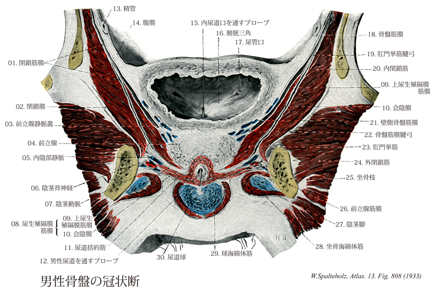
- 808_01【Obturator fascia閉鎖筋膜 Fascia obturatoria】 Stronger portion of parietal pelvic fascia covering the obturator internus.
→(壁側骨盤筋膜の上部は内閉鎖筋の内面を被っている骨盤筋膜のとくに強い部分。)
- 808_02【Obturator membrane閉鎖膜 Membrana obturatoria】 Membrane that closes the obturator foramen except for a superomedial opening that forms a portion of the obturator canal. Origin of the obturator intemus and externus muscles.
→(閉鎖膜は閉鎖孔の縁から起こって閉鎖管を除いた大部分を閉鎖する靱帯性の薄膜で、おもに横走する線維束よりなる。閉鎖膜の内外両面は、周囲の骨とともにそれぞれ内閉鎖筋および外閉鎖筋の起始となる。)
- 808_03【Prostatic venous plexus♂前立腺静脈叢;陰部静脈叢(♂) Plexus venosus prostaticus; Plexus venosus pudendalis♂】 Venous plexus around the prostate that is connected with the adjacent vesical venous plexus.
→(前立腺静脈叢は恥骨結合の下縁から前立腺の前面、さらにその外側部の結合組織のなかに発達した静脈叢で、これには前方から恥骨結合の下縁を通って深陰茎背静脈が流入するほか、膀胱下面および前立腺の静脈もそそぐ。膀胱静脈叢と連結して内腸骨静脈にそそぐほか、外腸骨静脈とも交通する。)
- 808_04【Prostate; Prostate gland; Prostatic glands前立腺 Prostata】 Chestnut-sized tubuloalveolar gland below the urinary bladder and having a smooth surface. It surrounds the urethra.
→(前立腺は膀胱底の下に密接し、骨盤底の上にのる30~50個の胞状管状腺である。男性の尿道起始部を取り囲んで栗の実に似た形をしている。強靱な結合組織の被膜に被われる。外周に近い部部分は色がやや暗く見え(いわゆる外腺exogland)、中心部は白っぽい(いわゆる内腺endogland)。Prostataはpro(前に)stata(立つもの)という意味である。古代ギリシャでは永来ヒトの前に立って護衛する人をprostatesといったそうである。前立腺を初めて記載し、prostatesと名付けたのは、アレキサンドリア学派のエラシストラトスErasistratus(BC310-250)である。前立腺が膀胱の前に立ってこれを守っていると考えたのだろう。日本では、以前には摂護腺と呼ばれていたが、ギリシャ語の直訳の前立腺が現在では正規の用語になっている。)
- 808_05【Internal pudendal vein内陰部静脈 Vena pudenda interna】 Vein running within the lateral wall of the ischiorectal fossa and passing through the lower portion of the greater sciatic foramen into the pelvis.
→(内陰部静脈は内陰部動脈に伴行するが、主として陰茎(核)深静脈のつづきとしてはじまり、坐骨直腸窩の外側壁に沿って背側へ走り、仙結節靱帯と仙棘靱帯の間を経て、梨状筋下孔から骨盤腔に入り、内腸骨静脈へそそぐ。この間、外陰部、肛門よりの静脈を入れる。)
- 808_06【Dorsal nerve of penis♂陰茎背神経(♂) Nervus dorsalis penis♂】 Paired nerve lying on the dorsum of penis that also sends branches to its inferior aspect.
→(陰茎背神経は深会陰横筋に分枝したあと、これを貫いて陰茎または陰核背面に達し、亀頭、包皮、尿道粘膜などに分布する。)
- 808_07【Artery of penis陰茎動脈 Arteria penis】
→()
- 808_08【Fascia of urogenital diaphragm尿生殖隔膜筋膜 Fasciae diaphragmatis urogenitalis】
→(")
- 808_09【Superior fascia of urogenital diaphragm上尿生殖隔膜筋膜;内尿生殖隔膜筋膜 Fascia diaphragmatis urogenitalis superior; Fascia diaphragmatis urogenitalis interna】 Obsolete term. Current scientific opinion holds that there is no complete boundary to the deep perineal space.
→(深会陰横筋の坐骨直腸窩側にある筋膜。 (Feneis))
- 808_10【Inferior fascia of urogenital diaphragm; Perineal membrane下尿生殖隔膜筋膜;会陰膜;外尿生殖隔膜筋膜 Membrana perinei; Fascia diaphragmatis urogenitalis inferior; Fascia diaphragmatis urogenitalis externa】
→(深会陰横筋の前下方にある筋膜。 (Feneis))
- 808_11【Urethral sphincter muscle; Sphincter urethrae muscle尿道括約筋;隔膜尿道括約筋;膜様尿道括約筋 Musculus sphincter urethrae; Musculus sphincter urethrae diaphragmaticae; Musculus sphincter urethrae membranaceae】
→(尿道括約筋は深会陰横筋から概念的に区分される尿道膜を環状に取り巻く筋線維束である。尿道の前方で会陰膜からの表層線維束は反対側からの線維と織り合い、尿道の後方で会陰へ放散している。深層線維束は内陰部血管のまわりの結合組織から起こり、尿道の膜性部を取り囲む筋性の輪を形成している。一般の形態的および機能的特徴に基づいて、深会陰横筋と尿道括約筋はともに尿道収縮筋とみなされる。男性では尿道括約筋の骨格筋線維は深会陰横筋の赤筋線維より通常やや明調ではるかに薄いが、前立腺に入り込む。一方、女性では押送する線維は腟により尿道後方でさえぎられており、その壁もまた尿道括約筋から放散する線維、すなわちいわゆる腟括約筋(尿道腟括約筋)を受けている。経産婦では、深会陰横筋の大部分が結合組織線維束によりとって代わる。平滑筋線維束は深会陰横筋と尿道括約筋の両方に埋もれているので、尿道や前立腺や腟などの平滑筋組織と明確に区別できない。深会陰横筋と尿道括約筋は尿道の前面で互いに区別されない。両筋は尿道筋壁から深会陰横筋を被う結合組織へ、女性では外側に、男性では背側に放散する平滑筋束により尿道から隔離されている。)
- 808_12【Male urethra男性尿道 Urethra masculina】
→(男の尿道は長さ約15~20cmである。膀胱頚の内尿道口に始まり、前立腺内を走り、尿道生殖隔膜を貫通し、陰茎の体を通って亀頭の前端で外尿道口に開く。尿道は走行によって、前立腺部・隔膜部・海綿体部の3部に分けられる。まず前立腺を貫通し、これを尿道の前立腺部という。後(背)壁中央には尿道稜があり、膀胱垂の連続する縦の隆起である。尿同僚の中央部は紡錘状にふくらみ、これを精丘という。精丘には前立腺小室が盲嚢として開く。これは胎生期のMueller管の名ごりで、男性子宮または男性腟ともいう。前立腺小室の両側に射精管が開口し、これより先の尿道は尿路と精液の通路を兼ねる。精丘両側のへこみが前立腺洞で、多くの前立腺管が開口する。つづいて尿道は尿生殖隔膜を貫く。これが隔膜部で、約1cm長。さらに外尿道口までが海綿体部で、12~14cm長。亀頭内の部分が膨大し、ここを尿道腺窩という。その後端上壁に舟状窩弁とよばれるヒダがある。海綿体部の粘膜には陥凹が多数みられ、尿道腺(リットレ腺)が開口する。)
- 808_13【Ductus deferens; Deferent duct精管 Ductus deferens; Vas deferens】 The course of the ca. 50 cm long ductus deferens is initially tortuous, then becomes straight. It is a continuation of the duct of epididymis, opening into the urethra.
→(精巣上体からはじまる精巣の分泌管で、精巣上体尾につづく精子を送る通路。精索中にある。全長約30cm(延ばせばその2倍)、膀胱底で紡錘状に膨れ、精管膨大部といい、内部に膨大部憩室を含む。膨大部の下端で、精嚢が精嚢排出管を経て合流し、これより遠位では精管は射精管と呼ばれ、尿道前立腺部後壁にある精丘の上で、尿道に開く。)
- 808_14【Peritoneum腹膜 Peritoneum】
→(腹膜は中皮と不規則な結合組織の薄い層からなる漿膜で、腹壁の内面(横隔膜の下面・骨盤壁の内面も含む)を被い、さらに腹腔・骨盤腔にあるいろいろな臓器の表面を包む。腹壁の内面を被う腹膜を腹膜を壁側腹膜といい、臓器の表面を被う腹膜を臓側腹壁という。)
- 808_15【Internal urethral orifice内尿道口 Ostium urethrae internum; Orificium urethrae internum】 Opening to the urethra from the urinary bladder. Due to invagination of the uvula of bladder, its ventral aspect appears convex in cross-section.
→(膀胱三角の前尖端で尿道が始まるところ。 (Feneis))
- 808_16【Trigone of bladder; Bladder mucosa膀胱三角 Trigonum vesicae】 Triangular area between the openings of the ureters and the exit site of the urethra, with underlying ureter musculature. It is firmly attached to the mucous membrane and hence does not have any folds.
→(尿管の開口部と尿道の出口の間にはさまれた三角形の領域で、ここでは粘膜が筋層と緊密に結合しているので、ヒダが認められない。 (Feneis))
- 808_17Mercier's bar【Ureteric orifice; Ureteric opening尿管口 Ostium ureteris; Orificium ureteris】 Slitlike opening of the ureter.
→(尿管の開口部で裂目状。 (Feneis))
- 808_18【Pelvic fascia骨盤部の筋膜;骨盤筋膜 Fascia pelvis; Fascia pelvica】 Continuation of the transversalis fascia into the pelvis. It splits into a visceral layer covering the pelvic viscera and a parietal layer covering the wall of the pelvis.
→(骨盤筋膜は骨盤腔中の諸構造を被う結合組織の膜で、壁側骨盤筋膜と臓側骨盤筋膜とを分かち、前者は上方は骨盤入口の分界線から下方は仙結節靱帯、坐骨結節、坐骨枝、恥骨の下枝に至り、その前方部は男で恥骨前立腺靱帯、女で恥骨膀胱靱帯などをつくり、その両側のものの間に陰茎背動脈または陰核背動脈を通ずる。壁側骨盤筋膜の上部は内閉鎖筋の内面を被って閉鎖筋膜という。)
- 808_19【Tendinous arch of levator ani muscle肛門挙筋腱弓;閉鎖筋膜腱弓 Arcus tendineus musculi levatoris ani; Arcus tendineus fasciae obturatoriae】 Tendinous arch of variable thickness formed by the obturator fascia at the origin of the levator ani.
→(上骨盤隔膜筋膜は閉鎖筋膜と合する線において肛門挙筋腱弓をなして肛門挙筋の起点をつくり、また下骨盤隔膜筋膜と内外相応じて肛門挙筋と尾骨筋とを包み骨盤出口の大部分を閉ざすロート上の骨盤隔膜を形成する。)
- 808_20【Obturator internus muscle; Internal obturator muscle内閉鎖筋 Musculus obturator internus】 o: Internal surface of obturator membrane and surrounding area, i: Trochanteric fossa of greater trochanter. Lateral rotation, abduction, adduction. I: Sacral plexus.
→(内閉鎖筋と2つの双子筋は発生的にはひとまとまりである。内閉鎖筋はその起始を骨盤腔内へ移し、閉鎖膜上および閉鎖孔の骨性枠から起こるに至った。小坐骨孔縁(軟骨でおおわれている)が視点となり、内閉鎖筋包が介在し、ここで急に走行を骨盤外へ変える。骨盤の外にある部分は3分筋のうちの2頭、つまり上下の双子筋を多少なりともおおう。上双子筋は坐骨棘を発し、下双子筋は坐骨結節を発する。内閉鎖筋の停止腱の上下縁にはそれぞれ上下の双子筋が合流し、転子窩に終わる。骨盤内にある内閉鎖筋は強い内閉鎖筋膜に包まれ、これが肛門挙筋の起始となる。内閉鎖筋膜は肛門挙筋起始部では弓状をした腱様の筋膜束(肛門挙筋腱弓)で補強さえている。肛門挙筋腱弓よりも上で、内閉鎖筋は小骨盤の筋性壁をつくり、その筋膜は壁側骨盤筋膜の一部となる。これより下では内閉鎖筋とその筋膜は外側部において、骨盤底の下にある結合組織性の部位、すなわち坐骨直腸窩を区画する。)
- 808_21【Parietal pelvic fascia; Endopelvic fascia壁側骨盤筋膜;骨盤内筋膜 Fascia pelvis parietalis; Fascia endopelvina】
→(壁側骨盤筋膜は上方は骨盤入口の分界線から下方は仙結節靱帯、坐骨結節、坐骨枝、恥骨の下枝に至り、その前方部は男で恥骨前立腺靱帯、音亜出恥骨膀胱靱帯などをつくる。)
- 808_22【Tendinous arch of pelvic fascia骨盤筋膜腱弓 Arcus tendineus fasciae pelvis】 Tendinous thickening of pelvic fascia that passes in an arch from the pubic symphysis to the levator ani and continues posteriorly to the ischial spine. It corresponds to a band giving exit to the visceral vessels and nerves from the lateral pelvic wall and providing an especially firm attachment for the pelvic fascia to the pelvic wall. Formation of the puboprostatic and pubovesical ligaments.
→(上骨盤隔膜筋膜のうち恥骨結合から坐骨棘へ走る弓状筋性の肥厚を骨盤筋膜腱弓(古くは白板と呼んだ)という。この線に沿って、内臓の血管・神経が骨盤側壁へと走り、骨盤結合組織が骨盤壁ととくに強固に結合している。)
- 808_23【Levator ani muscle肛門挙筋 Musculus levator ani】 Principle muscle of the pelvic diaphragm. It is derived from the abdominal wall musculature and permeated by smooth-muscle cells. I: Sacral plexus, S2-S5. It consists of the following parts.
→(肛門挙筋の丈夫な前部(恥骨尾骨筋)は分界線直下の恥骨の内面から起こり、薄い後部(腸骨尾骨筋)は腸骨から起こる。その起始腱は内閉鎖筋筋膜に接して移行し、閉鎖筋膜から発する腱束を受ける。これらの線維の起始部では腱性の係留物(肛門挙筋腱弓)により強化されている。左右両側で恥骨尾骨筋の内側線維束は挙筋脚を形成している。それらの線維束は背方と尾方、また直腸の前では外側を通り、それぞれ会陰の中心腱へ放散する薄い前直腸線維束や前立腺挙筋として前立腺筋膜(あるいは恥骨腟筋として腟壁)へと分かれる。それより鼻側にある肛門挙筋の線維束は恥骨直腸筋として直腸の背側を取り囲み、反対側の線維と共にループを形成する。恥骨尾骨筋の外側束は尾骨と仙骨の背側に広がる。腸骨尾骨筋の筋線維は尾骨と仙骨に付き、また肛門と尾骨の間では強靱な線維束である肛門尾骨靱帯に付いている。)
- 808_24【Obturator externus muscle; External obturator muscle外閉鎖筋 Musculus obturator externus】 o: External surface of obturator membrane and surrounding area, i: Trochanteric fossa. Lateral rotation and adduction at the hip joint. I: Obturator nerve.
→(外閉鎖筋は閉鎖膜外面および閉鎖孔内下方の骨縁に起こる。腹内側へ走り、股関節の背側で大腿骨頚と頭をまわりこんでから錐状の停止腱に移行し、腹外側へ向かって転子窩に停止する。)
- 808_25【Ramus of ischium坐骨枝;坐骨下枝 Ramus; Ramus inferior (Os ischii)】 The part of the ischium below the obturator foramen. Its anterior end articulates with the inferior pubic ramus.
→(坐骨結節のところから前方に伸びて恥骨下枝に合する部で、閉鎖孔の下縁の後半をつくる。坐骨結節につづく下縁がやや厚く、閉鎖孔に向かう上縁が鋭い三角柱状をなす。)
- 808_26【Prostatic fascia前立腺筋膜 Fascia prostatae】
→(周囲と癒着している前立腺の筋膜。(Feneis))
- 808_27【Crus of penis陰茎脚;陰茎海綿体脚 Crus penis; Crura corporis cavernosi penis】 Projection from the corpus cavernosum of penis attached to the inferior pubic ramus.
→(陰茎海綿体と尿道海綿体とは陰茎体から近位側に向かってのび、それぞれ、陰茎脚および尿道球となり陰茎根をつくる。すなわち左右両側の陰茎海綿体は恥骨結合の下側で、陰茎脚となり、左右に分かれて恥骨弓に沿って走る。)
- 808_28【Ischiocavernosus muscle坐骨海綿体筋 Musculus ischiocavernosus】 Male: Muscle extending from the ramus of ischium over the cms of the penis to the tunica albuginea. Smaller bundles of muscle fibers run over the penis below the pubic symphysis to the contralateral side.
→(坐骨海綿体筋は男性より女性のほうが発達が弱い。この筋は坐骨枝より起こり陰核脚を被い、その腱性線維は外下表面に付く。その筋は陰核海綿体を圧し、血液を押し付け流出を妨げ、それにより陰核の勃起成立を助ける。)
- 808_29【Bulbospongiosus muscle球海綿体筋 Musculus bulbospongiosus; Musculus bulbocavernosus】 Male: Muscle arising from the perineal body and the inferior aspect of the corpus spongiosum of penis, passing to the perineal membrane and dorsum of penis. It is unpaired. It acts to compress the bulb of penis and transport urethral contents further. ABC Female: Muscle that originates on the ramus of ischium, attaching to and covering the cms of clitoris. It assists in filling the cavernous bodies with blood. I: Pudendal nerve.
→(男性では球海綿体は尿道球の周辺を不体の筋として回るが、会陰の中心腱と尿道海綿体下側の正中縫線から起こる。球海綿体は前方へ放散し、海綿体のまわり下尿生殖隔膜筋膜や尿道海綿体へ向かい、また前筋線維をもって陰茎背部へ付く。この筋は随意的または反射的に尿道球を圧迫し、それにより尿道の内容を駆出する。女性では球海綿体筋は男性のように全長で1つの筋にはなっていない。2つの筋が会陰の中心腱より起こるが、各筋はそれぞれ引き続き前庭球と大前庭腺を被っている。その筋束は前庭球や陰核海綿体に停止し、陰核体後部で反対側からの筋線維と絡み合っている。この筋は大前庭腺を反射的に空にし、血液を前庭球の後方拡大部から送り出し、またオルガスムの際外腟口を収縮させる。)
- 808_30【Bulb of penis; Bulbus portion of spongy urethra尿道球;陰茎球;尿道海綿体球 Bulbus penis; Bulbus urethrae; Bulbus corporis cavernosi urethrae】 Posterior, thickened end of the corpus spongiosum.
→(陰茎球は左右の陰茎脚の間で後上方に向かって球状に肥大したもので、尿生殖隔膜の下面(会陰膜)に付着する。)